Identification and clonal characterisation of a progenitor cell sub-population in normal human articular cartilage
- PMID: 20976230
- PMCID: PMC2954799
- DOI: 10.1371/journal.pone.0013246
Identification and clonal characterisation of a progenitor cell sub-population in normal human articular cartilage
Abstract
Background: Articular cartilage displays a poor repair capacity. The aim of cell-based therapies for cartilage defects is to repair damaged joint surfaces with a functional replacement tissue. Currently, chondrocytes removed from a healthy region of the cartilage are used but they are unable to retain their phenotype in expanded culture. The resulting repair tissue is fibrocartilaginous rather than hyaline, potentially compromising long-term repair. Mesenchymal stem cells, particularly bone marrow stromal cells (BMSC), are of interest for cartilage repair due to their inherent replicative potential. However, chondrocyte differentiated BMSCs display an endochondral phenotype, that is, can terminally differentiate and form a calcified matrix, leading to failure in long-term defect repair. Here, we investigate the isolation and characterisation of a human cartilage progenitor population that is resident within permanent adult articular cartilage.
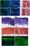
Methods and findings: Human articular cartilage samples were digested and clonal populations isolated using a differential adhesion assay to fibronectin. Clonal cell lines were expanded in growth media to high population doublings and karyotype analysis performed. We present data to show that this cell population demonstrates a restricted differential potential during chondrogenic induction in a 3D pellet culture system. Furthermore, evidence of high telomerase activity and maintenance of telomere length, characteristic of a mesenchymal stem cell population, were observed in this clonal cell population. Lastly, as proof of principle, we carried out a pilot repair study in a goat in vivo model demonstrating the ability of goat cartilage progenitors to form a cartilage-like repair tissue in a chondral defect.
Conclusions: In conclusion, we propose that we have identified and characterised a novel cartilage progenitor population resident in human articular cartilage which will greatly benefit future cell-based cartilage repair therapies due to its ability to maintain chondrogenicity upon extensive expansion unlike full-depth chondrocytes that lose this ability at only seven population doublings.
Conflict of interest statement
Figures


Similar articles
-
Clonal chondroprogenitors maintain telomerase activity and Sox9 expression during extended monolayer culture and retain chondrogenic potential.Osteoarthritis Cartilage. 2009 Apr;17(4):518-28. doi: 10.1016/j.joca.2008.08.002. Epub 2008 Nov 17. Osteoarthritis Cartilage. 2009. PMID: 19010695
-
Articular Cartilage Repair with Mesenchymal Stem Cells After Chondrogenic Priming: A Pilot Study.Tissue Eng Part A. 2018 May;24(9-10):761-774. doi: 10.1089/ten.TEA.2017.0235. Epub 2017 Nov 30. Tissue Eng Part A. 2018. PMID: 28982297
-
Isolation and characterisation of nasoseptal cartilage stem/progenitor cells and their role in the chondrogenic niche.Stem Cell Res Ther. 2020 May 14;11(1):177. doi: 10.1186/s13287-020-01663-1. Stem Cell Res Ther. 2020. PMID: 32408888 Free PMC article.
-
Reserve or Resident Progenitors in Cartilage? Comparative Analysis of Chondrocytes versus Chondroprogenitors and Their Role in Cartilage Repair.Cartilage. 2018 Apr;9(2):171-182. doi: 10.1177/1947603517736108. Epub 2017 Oct 19. Cartilage. 2018. PMID: 29047310 Free PMC article.
-
Cell-based articular cartilage repair: the link between development and regeneration.Osteoarthritis Cartilage. 2015 Mar;23(3):351-62. doi: 10.1016/j.joca.2014.11.004. Epub 2014 Nov 11. Osteoarthritis Cartilage. 2015. PMID: 25450846 Free PMC article. Review.
Cited by
-
Strategies to Convert Cells into Hyaline Cartilage: Magic Spells for Adult Stem Cells.Int J Mol Sci. 2022 Sep 22;23(19):11169. doi: 10.3390/ijms231911169. Int J Mol Sci. 2022. PMID: 36232468 Free PMC article. Review.
-
Comparison of Electrophysiological Properties and Gene Expression between Human Chondrocytes and Chondroprogenitors Derived from Normal and Osteoarthritic Cartilage.Cartilage. 2020 Jul;11(3):374-384. doi: 10.1177/1947603518796140. Epub 2018 Aug 23. Cartilage. 2020. PMID: 30139266 Free PMC article.
-
Articular Cartilage-From Basic Science Structural Imaging to Non-Invasive Clinical Quantitative Molecular Functional Information for AI Classification and Prediction.Int J Mol Sci. 2023 Oct 7;24(19):14974. doi: 10.3390/ijms241914974. Int J Mol Sci. 2023. PMID: 37834422 Free PMC article. Review.
-
Pathomechanisms of Posttraumatic Osteoarthritis: Chondrocyte Behavior and Fate in a Precarious Environment.Int J Mol Sci. 2020 Feb 25;21(5):1560. doi: 10.3390/ijms21051560. Int J Mol Sci. 2020. PMID: 32106481 Free PMC article. Review.
-
Cell Interplay in Osteoarthritis.Front Cell Dev Biol. 2021 Aug 3;9:720477. doi: 10.3389/fcell.2021.720477. eCollection 2021. Front Cell Dev Biol. 2021. PMID: 34414194 Free PMC article. Review.
References
-
- Khan IM, Redman SN, Williams R, Dowthwaite GP, Oldfield SF, et al. The development of synovial joints. Curr Top Dev Biol. 2007;79:1–36. - PubMed
-
- Cournil-Henrionnet C, Huselstein C, Wang Y, Galois L, Mainard D, et al. Phenotypic analysis of cell surface markers and gene expression of human mesenchymal stem cells and chondrocytes during monolayer expansion. Biorheology. 2008;45:513–526. - PubMed
-
- Schnabel M, Marlovits S, Eckhoff G, Fichtel I, Gotzen L, et al. Dedifferentiation-associated changes in morphology and gene expression in primary human articular chondrocytes in cell culture. Osteoarthritis Cartilage. 2002;10:62–70. - PubMed
-
- Dell'Accio F, De Bari C, Luyten FP. Molecular markers predictive of the capacity of expanded human articular chondrocytes to form stable cartilage in vivo. Arthritis Rheum. 2001;44:1608–1619. - PubMed
-
- Benya PD, Shaffer JD. Dedifferentiated chondrocytes reexpress the differentiated collagen phenotype when cultured in agarose gels. Cell. 1982;30:215–224. - PubMed
Publication types
MeSH terms
Substances
LinkOut - more resources
Full Text Sources
Other Literature Sources
Medical

