Case Reports
. 2010 Oct 12;5(10):e13315.
doi: 10.1371/journal.pone.0013315.

A systematic molecular pathology study of a laboratory confirmed H5N1 human case

Affiliations
Case Reports

A systematic molecular pathology study of a laboratory confirmed H5N1 human case

Rongbao Gao et al. PLoS One. .

Abstract

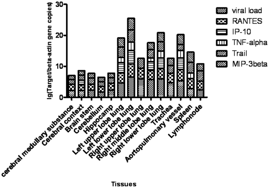
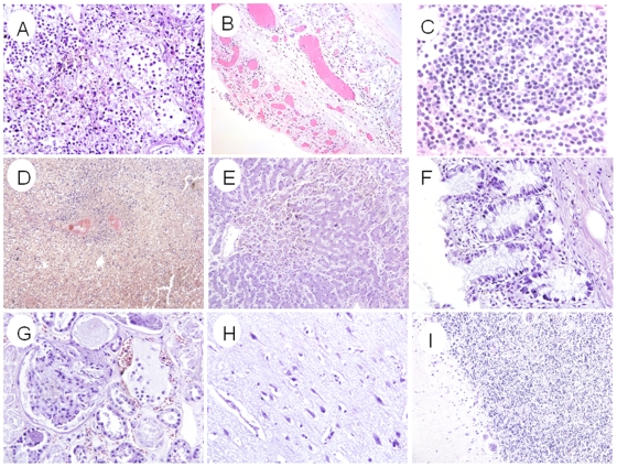
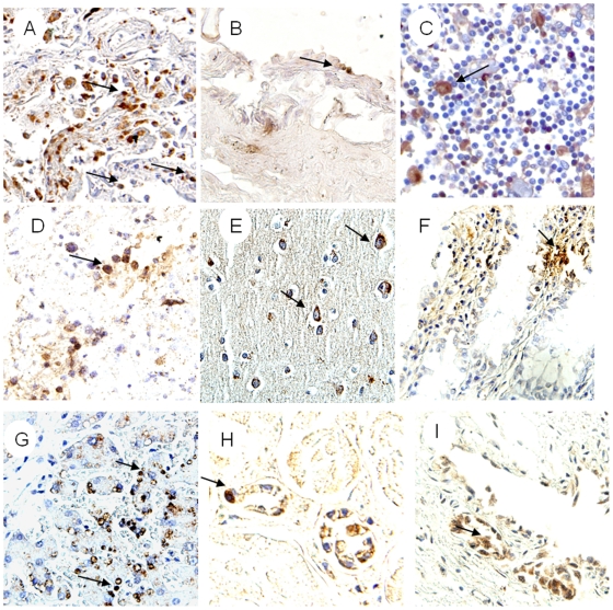
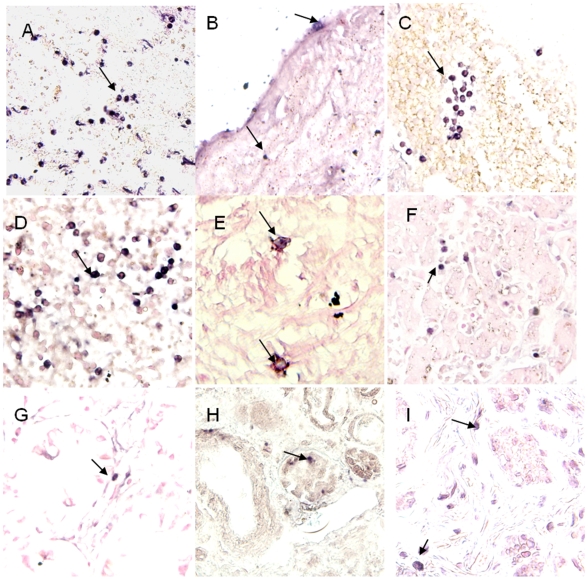
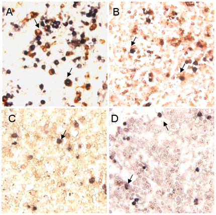
Autopsy studies have shown that human highly pathogenic avian influenza virus (H5N1) can infect multiple human organs other than just the lungs, and that possible causes of organ damage are either viral replication and/or dysregulation of cytokines and chemokines. Uncertainty still exists, partly because of the limited number of cases analysed. In this study, a full autopsy including 5 organ systems was conducted on a confirmed H5N1 human fatal case (male, 42 years old) within 18 hours of death. In addition to the respiratory system (lungs, bronchus and trachea), virus was isolated from cerebral cortex, cerebral medullary substance, cerebellum, brain stem, hippocampus ileum, colon, rectum, ureter, aortopulmonary vessel and lymph-node. Real time RT-PCR evidence showed that matrix and hemagglutinin genes were positive in liver and spleen in addition to positive tissues with virus isolation. Immunohistochemistry and in-situ hybridization stains showed accordant evidence of viral infection with real time RT-PCR except bronchus. Quantitative RT-PCR suggested that a high viral load was associated with increased host responses, though the viral load was significantly different in various organs. Cells of the immunologic system could also be a target for virus infection. Overall, the pathogenesis of HPAI H5N1 virus was associated both with virus replication and with immunopathologic lesions. In addition, immune cells cannot be excluded from playing a role in dissemination of the virus in vivo.


Conflict of interest statement

Competing Interests: The authors have declared that no competing interests exist.

Figures

Figure 1
Figure 1. The correlation between viral load and levels of proinflammatory factors in various tissues.
Similar with viral load assay, RANTES, IP-10, TNF-α, TRAIL and MIP-3β genes in respiratory tissues, brains, lymph-node and spleen were quantified by real-time RT-PCR. The ratios between proinflammatory factors and β-actin gene copies were presented by logarithm.
Figure 2
Figure 2. The histopathologic features by H&E stain.
(A) Lung tissue showed diffuse alveolar damage characterized by intraalveolar edema, mononuclear inflammation cell infiltration, necrosis of alveolar lining cells, desquamation of pneumocytes in alveolar spaces and hyaline membrane formation. (B) Trachea showed focal denudation of the epithelium with edema and mononuclear inflammatory cell infiltrates. (C) Lymph node was congested with depletion of lymphocytes. (D) Spleen showed depletion of lymphocytes with congestion and organized infarcts. (E) Liver was congested with edema and focal fatty degeneration. (F) Intestine showed mild inflammatory infiltrates in lamina propria. (G) Kidney was congested with edema. (H) Cerebral cortex showed edema with focal neuronal necrosis in hippocampus area. (I) Cerebellum showed focal dilatation between Purkinje cells layer and particle cells layer. Original magnifications: (A–C)×20, (D) ×10, (E–I) ×20.
Figure 3
Figure 3. The results of IHC stain with NP against antibody.
Positive stain seen with diaminobenzidine (brown; Dako). Slides counterstained with haematoxylin. (A) Positive stain presented in pneumocytes of lung (arrow). (B) Endotheliocyte of aortopulmonary vessel (arrow). (C) Positive stain in nuclei and cytoplasm of axillary lymphoid-node (arrow). (D) Positive stain in lymphocytes of spleen (arrow). (E) Positive stain in cytoplasm of neuron and gliocyte from cerebral cortex (arrows). (F) Positive stain in nuclei and cytoplasm of intestine mucosa (arrows). (G) Positive stain in nuclei of mononuclear-like cells (arrows) with morphological features of macrophages from liver. (H) Positive stain in cytoplasm and nuclei of endotheliocytes of renal contex. (I) Positive stain in nuclei and cytoplasm of epithelial cells from ureter (arrow). Original magnifications: (A) ×20, (B–E) ×40, (F–G) ×20, (H–I) ×40.
Figure 4
Figure 4. The results of ISH stain with probes of NP gene and HA gene.
Positive stain seen with nitroblue tetrazolium/5-bromo-4-choloro-3-indolyl phosphate (purple-blue, Roch). Slides counterstained with nucleic fast red solution. (A) Positive stain (with NP sense probe) in nuclear and cytoplasm of pneumocytes (arrows). (B) Positive stain (with NP antisense probe) in nuclei and cytoplasm of Endotheliocyte from aortopulmonary (arrows). (C) Positive stain (with NP antisense probe) in lymphocytes of oxter lymph-node (arrows). (D) Positive stain (with NP antisense probe) in lymphocytes and monocytes from spleen (arrows). (E) Positive stain (with NP antisense probe) in nuclei and cytoplasm of neuron from Cerebrum cortex (arrow). (F) Positive stain (with NP antisense probe) in cytoplasm of epithelial cells from Colon (arrow). (G) Positive stain (with NP antisense probe) in lymphocytes of liver (arrow). (H) Positive stain (with HA antisense probe) in cytoplasm of endotheliocyte of renal contex (arrow). (I) Positive stain (with NP antisense probe) in nuclei of epithelial cells of ureter. Original magnifications: (A) ×20, (B–I) ×40.
Figure 5
Figure 5. Double-labeled stain combined ISH for HA gene and IHC for the cell superfic markers of macrophage CD68 protein, T lymphocyte CD3 protein, progenitor cells CD34 protein and follicular dendritic cells CD35 protein.
Positive of in-situ hybridization seen with nitroblue tetrazolium/5-bromo-4-choloro-3-indolyl phosphate (purple-blue, Roch), IHC with diaminobenzidine (brown; Dako). Slides counterstained with 2% nuclear fast red solution. (A) Double positive cells in lung. The viral sequence presented in nucleic and cytoplasm of CD68+ cell (arrow). Viral sequence presented in nucleic and cytoplasm what showed the feature of endocytosis. (B) Viral sequence positive stain presented in CD3+ T lymphocytes of spleen (arrow). (C) Viral sequence positive signal presented in CD34+ cell of spleen (arrow). (D) Viral sequence positive stain presented in CD35+ cell of spleen (arrow). Original magnifications: (A–D) ×40.

References

    1. Cox NJ, Subbarao K. Global epidemiology of influenza: past and present. Annu Rev Med. 2000;51:407–21. - PubMed
    1. Webster RG, Bean WJ, Gorman OT, Chambers TM, Kawaoka Y. Evolution and ecology of influenza A viruses. Microbiol Rev. 1992;56(1):152–79. - PMC - PubMed
    1. Subbarao K, Klimov A, Katz J, Regnery H, Lim W, et al. Characterization of an avian influenza A (H5N1) virus isolated from a child with a fatal respiratory illness. Science. 1998;279(5349):393–6. - PubMed
    1. World Health Organization. 2009. Cumulative number of confirmed human cases of avian influenza A/(H5N1) reported to who. Available at: http://www.who.int/csr/disease/avian_influenza/country/cases_table_2009_....
    1. Ungchusak K, Auewarakul P, Dowell SF, Kitphati R, Auwanit W, et al. Probable person-to-person transmission of avian influenza A (H5N1). N Engl J Med. 2005;352(4):333–40. - PubMed

Publication types