. 2011 May;68(10):1785-98.
doi: 10.1007/s00018-010-0546-9. Epub 2010 Oct 26.

Delayed c-Fos activation in human cells triggers XPF induction and an adaptive response to UVC-induced DNA damage and cytotoxicity

Affiliations

Delayed c-Fos activation in human cells triggers XPF induction and an adaptive response to UVC-induced DNA damage and cytotoxicity

Maja T Tomicic et al. Cell Mol Life Sci. 2011 May.

Abstract

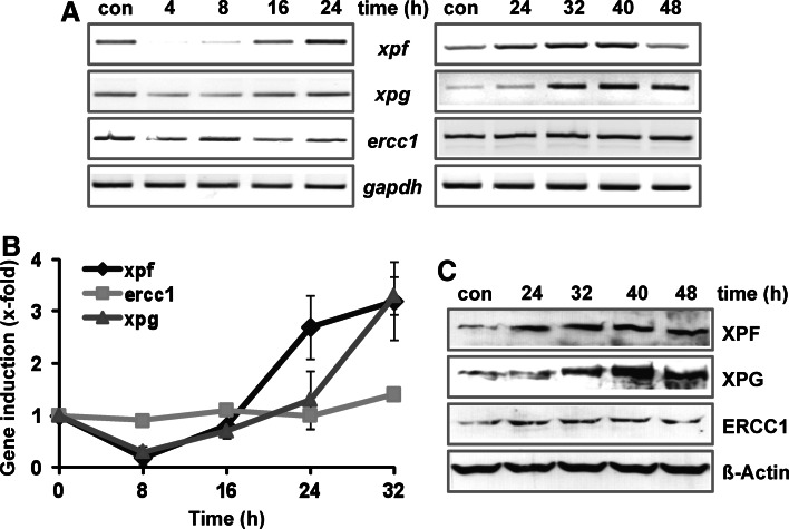
The oncoprotein c-Fos has been commonly found differently expressed in cancer cells. Our previous work showed that mouse cells lacking the immediate-early gene c-fos are hypersensitive to ultraviolet (UVC) light. Here, we demonstrate that in human diploid fibroblasts UV-triggered induction of c-Fos protein is a delayed and long-lasting event. Sustained upregulation of c-Fos goes along with transcriptional stimulation of the NER gene xpf, which harbors an AP-1 binding site in the promoter. Data gained on c-Fos knockdown and c-Fos overexpressing human cells provide evidence that c-Fos/AP-1 stimulates upregulation of XPF, thereby increasing the cellular repair capacity protecting from UVC-induced DNA damage. When these cells are pre-exposed to a low non-toxic UVC dose and challenged with a subsequent high dose of UVC irradiation, they show accelerated repair of UVC-induced DNA adducts and reduced cell kill. The data indicate a protective role of c-Fos induction by triggering an adaptive response pathway.


Conflict of interest statement

The authors declare that there is no conflict of interest.

Figures

Fig. 1
Fig. 1
UVC-induced activation of the MAPK cascade and induction of Fos proteins. a Exponentially growing VH10tert cells were exposed to 10 J/m2 UVC. At different time points after exposure, total RNA was isolated and semi-quantitative RT-PCR was performed using c-fos, fosB, fra1 or, as loading control, gapdh specific primers. b, c Exponentially growing VH10tert cells were exposed to 10 J/m2 UVC for the indicated times. Protein extracts were prepared and subjected to Western blot analysis. The filter was incubated with c-Fos, FosB or Fra1 specific antibodies (b) or incubated with p-JNK, JNK, p-p38K, p38K, p-ERK1/2 and ERK1/2 specific antibodies (c); the two phosphorylated JNK specific bands (JNK1 p46 and JNK2 p54) are labeled by arrows. d Left panel: Exponentially growing VH10tert cells were exposed to 10 J/m2 UVC; 16 h later 10 μM of a specific inhibitor for JNK1/2/3 (SP600125), p38K (SB103580), and MEK1/2 (UO126), respectively, was added. Additional 8 h later, protein extracts were prepared and subjected to Western blot analysis. The filter was incubated with c-Fos and ERK2 specific antibodies. d Right panel: Exponentially growing VH10tert cells were pre-incubated for 1 h with a specific inhibitor for JNK, p38K and MEK1/2, respectively. Thereafter cells were exposed to 10 J/m2 UVC, and the conditioned medium containing the inhibitor was re-added. Then 24 h later protein extracts were prepared and subjected to Western blot analysis. The filter was incubated with c-Fos and ERK2 specific antibodies
Fig. 2
Fig. 2
UVC-mediated induction of NER proteins. a Exponentially growing VH10tert cells were exposed to 10 J/m2 UVC. At different time points after exposure, total RNA was isolated and semi-quantitative RT-PCR was performed using xpf, xpg, ercc1 or, as loading control, gapdh specific primers. b Exponentially growing VH10tert cells were exposed to 10 J/m2 UVC for the indicated times. Total RNA was isolated and real-time RT-PCR was performed using xpf, xpg, ercc1 or, as loading control, gapdh specific primers. For quantification, the expression was normalized to gapdh and the untreated control was set to 1. Data are the mean of three independent experiments ± SD. c Exponentially growing VH10tert cells were exposed to 10 J/m2 UVC for the indicated time points. Protein extracts were prepared and subjected to Western blot analysis. The filter was incubated with XPF, XPG or ERCC1 specific antibodies. β-actin, loading control
Fig. 3
Fig. 3
Effect of c-Fos knockdown on XPF induction and NER capacity. a Exponentially growing VH10tert cells were transfected with c-Fos specific siRNA (si) or nonsense-siRNA (ns-si) and 24 h later exposed to 10 J/m2 UVC. Protein extracts were prepared 24 or 48 h after exposure and subjected to Western blot analysis. The filter was incubated with c-Fos specific antibodies. β-actin was used as loading control. b Exponentially growing VH10tert cells were transfected with c-Fos specific siRNA (si) or nonsense-siRNA (ns-si) and 24 h later exposed to 10 J/m2 UVC. Protein extracts were prepared 32 h after exposure and subjected to Western blot analysis. The filter was incubated with XPF or XPG specific antibodies. β-actin was used as loading control. The induction factor (IF) is derived from densitometric measurement of XPF and XPG signal and normalized to β-actin expression. c, d To analyze NER capacity upon c-Fos knockdown VH10tert cells were transfected with c-Fos specific siRNA (si) or nonsense-siRNA (ns-si) and 24 h later exposed to 10 J/m2 UVC. At different time points following irradiation, genomic DNA was isolated; equal amounts of DNA were blotted and subjected to incubation with c anti-CPD antibodies or d anti-(6-4)PP antibodies (con, non-exposed control). Representative blots are shown (left panel) and the mean data of three independent experiments ± SD (right panel). The CPD signal determined 5 min after treatment was set to 100%. Data were compared statistically using Student’s t test (**p < 0.01)
Fig. 4
Fig. 4
Effect of c-Fos knockdown on sensitivity to UVC light. To monitor the impact of c-Fos knockdown on sensitivity to UVC light, VH10tert cells were transfected with c-Fos specific siRNA (c-fos-si) or nonsense-siRNA (non-si); 48 h later, cells were irradiated with 10 (a) or 25 J/m2 UVC (b) and incubated for 96 or 144 h. Cells were stained with PI, and the sub-G1 fraction (apoptotic cells) was determined by flow cytometry. Experiments were repeated at least three times, mean values ± SD are shown, and data were compared statistically using Student’s t test (*p < 0.05, **p < 0.01)
Fig. 5
Fig. 5
Effect of c-Fos overexpression on XPF induction, NER capacity and apoptosis. a Human GM637 fibroblasts were transfected with a c-Fos expression plasmid. Parental GM637 cells and five clones expressing different amounts of c-Fos were selected. Total RNA and protein extracts were prepared from the individual clones and subjected to RT-PCR (upper panel) or Western blot analysis (lower panel). The expression of c-Fos was analyzed via c-Fos specific primers or antibodies. For loading control, gapdh or β-actin was detected. b Total RNA was prepared from the individual clones and subjected to RT-PCR. The expression of xpf and xpg was analyzed via specific primers. For loading control, gapdh was detected. c Exponentially growing clones were exposed to 10 J/m2 UVC. Total RNA was isolated at different time points after exposure and real-time RT-PCR was performed using xpf (left panel) or xpg (right panel) specific primers. For quantification, the expression was normalized to gapdh and the untreated control was set to 1. d To analyze NER capacity upon c-Fos overexpression, the different clones were exposed to 10 J/m2 UVC. At different time points following irradiation, genomic DNA was isolated, and equal amounts of DNA were blotted and incubated with anti-CPD antibodies (con, non-exposed control). e To monitor the impact of c-Fos overexpression on sensitivity to UVC light, the different cell clones were irradiated with 10 J/m2, and 96 h later cells were stained with PI and the sub-G1 fraction (apoptotic cells) was determined by flow cytometry. Experiments were repeated at least three times, mean values ± SD are shown, and data were compared statistically using Student’s t test (*p < 0.05, **p < 0.01)
Fig. 6
Fig. 6
Identification of AP-1 binding sites within the xpf promoter. a Oligonucleotides containing either the AP-1 binding site of the collagenase promoter (mmp1) or the AP-1 binding sites of the xpf promoter were incubated with nuclear extracts from VH10tert cells exposed to 10 J/m2 UVC light and harvested at different time points later and subjected to EMSA. The specific complex is indicated by an arrow. b For verifying the specificity of the reaction, a 5-, 10-, or 25-fold excess of unlabeled oligonucleotides containing either the p53 binding site of p21 (p21) or the AP-1 binding site of the collagenase promoter (mmp1) was added to the assay using labeled xpf promoter specific oligonucleotides. In addition EMSA was performed using point-mutated oligonucleotides (containing two exchanges in the xpf specific AP-1 binding site; xpf-mut) and nuclear extracts from VH10tert cells exposed to 10 J/m2 UVC light and harvested 8 and 24 h later. c EMSA supershift assay. Composition of the AP-1 factor bound to the mmp1 AP-1 and xpf AP-1 oligonucleotide was analyzed by the addition of specific antibodies against p53, c-Jun, or c-Fos to the reaction. The supershifted complex is indicated by an arrow
Fig. 7
Fig. 7
Adaptive response to UVC-induced damage. a, b VH10tert cells were either not pre-exposed (control) or pre-exposed to 5 J/m2 UVC. Thirty-two hours later the cells were exposed to 10 J/m2 UVC. At different time points later, genomic DNA was isolated, and equal amounts of DNA were blotted and subjected to incubation with anti-CPD (a) or anti-(6-4)PP antibodies (b) (con, non-exposed control). The representative experiment is shown in the left panel. The right panel shows the mean remaining CPDs in three independent experiments ± SD. c To monitor the impact of pre-exposure on sensitivity to UVC light, VH10tert cells were either pre-exposed or not pre-exposed to 5 J/m2 UVC and 32 h later exposed to 25 J/m2 UVC. One hundred twenty hours later, cells were stained with PI, and the sub-G1 fraction (apoptotic cells) was determined by flow cytometry. In addition, cells were irradiated with 5 J/m2 and incubated for 152 h. Experiments were repeated at least three times, mean values ± SD are shown, and data were compared statistically using Student’s t test (*p < 0.05)
Fig. 8
Fig. 8
Model of MAPK, c-Fos, and XPF expression under physiological and c-Fos overexpressing conditions upon UVC exposure. In normal unexposed fibroblasts under physiological conditions (upper flow chart) neither c-fos mRNA nor c-Fos protein is expressed. Upon UVC exposure MAPKs are activated and phosphorylate various transcription factors such as Elk1 and c-Jun, which are responsible for the transcriptional activation of c-fos mRNA. This is followed by expression of c-Fos protein, which in turn becomes activated via phosphorylation by MAPKs and binds to its partner, e.g., c-Jun, to form the active AP-1 complex, which finally activates the xpf promoter. In UVC-irradiated c-Fos overexpressing cells (lower flow chart) the first steps, namely induction of xpf mRNA and XPF protein, have already occurred because the MAPKs directly activate c-Fos leading to accelerated induction of XPF and faster repair of CPDs. Although overexpressed in unexposed cells, c-Fos is not transcriptionally active because it needs activation by MAPKs and a binding partner such as c-Jun. Following UVC exposure these proteins are activated, c-Fos containing AP-1 is formed, and target genes including xpf are induced

Similar articles

Cited by

References

    1. Christmann M, Tomicic MT, Roos WP, Kaina B. Mechanisms of human DNA repair: an update. Toxicology. 2003;193:3–34. doi: 10.1016/S0300-483X(03)00287-7. - DOI - PubMed
    1. Hoeijmakers JH. DNA damage, aging, and cancer. N Engl J Med. 2009;361:1475–1485. doi: 10.1056/NEJMra0804615. - DOI - PubMed
    1. Christmann M, Fritz G, Kaina B (2007) Induction of DNA repair genes in mammalian cells in response to genotoxic stress. In: D. Lankenau (Ed) Genome dynamics and stability, vol 1. Springer, Berlin, pp 383–398
    1. Adimoolam S, Ford JM. p53 and DNA damage-inducible expression of the xeroderma pigmentosum group C gene. Proc Natl Acad Sci USA. 2002;99:12985–12990. doi: 10.1073/pnas.202485699. - DOI - PMC - PubMed
    1. Montecucco A, Savini E, Biamonti G, Stefanini M, Focher F, Ciarrocchi G. Late induction of human DNA ligase I after UV-C irradiation. Nucleic Acids Res. 1995;23:962–966. doi: 10.1093/nar/23.6.962. - DOI - PMC - PubMed

Publication types

Substances

LinkOut - more resources