. 2011 Feb 7;8(1):143-52.
doi: 10.1021/mp100203a. Epub 2010 Dec 8.

Modulation of CD4+ T lymphocyte lineage outcomes with targeted, nanoparticle-mediated cytokine delivery

Affiliations

Modulation of CD4+ T lymphocyte lineage outcomes with targeted, nanoparticle-mediated cytokine delivery

Jason Park et al. Mol Pharm. .

Abstract

Within the immune system there is an exquisite ability to discriminate between "self" and "non-self" that is orchestrated by antigen-specific T lymphocytes. Genomic plasticity enables differentiation of naive CD4+ T lymphocytes into either regulatory cells (Treg) that express the transcription factor Foxp3 and actively prevent autoimmune self-destruction or effector cells (Teff) that attack and destroy their cognate target. An example of such plasticity is our recent discovery that leukemia inhibitory factor (LIF) supports Treg maturation in contrast to IL-6, which drives development of the pathogenic Th17 effector phenotype. This has revealed a LIF/IL6 axis in T cell development which can be exploited for modulation using targeted cytokine delivery. Here we demonstrate that LIF-loaded nanoparticles (NPs) directed to CD4+ T cells (i) oppose IL6-driven Th17 development; (ii) prolong survival of vascularized heart grafts in mice; and (iii) expand FOXP3+ CD4+ T cell numbers in a non-human primate model in vitro. In contrast, IL-6 loaded nanoparticles directed to CD4+ T cells increase Th17 development. Notably, nanoparticle-mediated delivery was demonstrated to be critical: unloaded nanoparticles and soluble LIF or IL-6 controls failed to recapitulate the efficacy of cytokine-loaded nanoparticles in induction and/or expansion of Foxp3+ cells or Th17 cells. Thus, this targeted nanoparticle approach is able to harness endogenous immune-regulatory pathways, providing a powerful new method to modulating T cell developmental plasticity in immune-mediated disease indications.


Conflict of interest statement

Competing Interests Statement

The authors declare no competing financial interests.

Figures

Figure 1
Figure 1. Nanotherapy-mediated modulation of CD4+ T cell differentiation
(A) Schematic model of cytokine-loaded, antibody (anti-CD4)-targeted PLGA nanoparticle. Avidin groups on the nanoparticle surface facilitate attachment of biotinylated targeting antibodies. Hydrolyis of the polymeric matrix releases entrapped, bioactive cytokine in sustained fashion. (B) Prior to in vitro stimulation and in vivo lymphocyte transfusion experiments (Figs. 2–4), nanoparticles were attached to CD4+ T cells via anti-CD4 antibodies. Nanoparticles encapsulating LIF (LIF-nano) were found to enhance Foxp3 expression, while nanoparticles encapsulating IL-6 (IL-6-nano) enhanced RORγT expression.
Figure 2
Figure 2. Characterization of cytokine-loaded PLGA nanoparticles
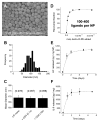
(A) Scanning electron micrograph (SEM) of PLGA nanoparticles. (B) Representative size distribution of PLGA nanoparticles based on analysis of SEM data using Scion image processing software. The mean particle diameter was 100 ± 20 nm (mean ± s.d. of n=3 batches). (C) Effect of ligand attachment on mean effective hydrodynamic diameter, overall sample polydispersity is reported in parentheses above the column. Data represent mean ± S.D. of 10 measurements. (D) Ligand attachment was quantified by measuring NP uptake of biotin-R-PE. LIF-nano were found to bind up to 1.6 ± 0.6 × 10¹³ molecules of biotin-R-PE per mg NP. Particle counts were between 5×10¹⁰ and 1×10¹¹NPs/mg. (E) Cumulative release of LIF or (F) IL-6 from PLGA nanoparticles in pg cytokine per milligram nanoparticles. Cytokine concentration was measured by ELISA and data represent mean ± s.d. (n=3 individual samples per time point).
Figure 3
Figure 3. Nanoparticle-delivered LIF and IL-6 have counter-regulatory effects on Foxp3 expression in vitro
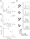
LIF supports Foxp3 expression following activation in the presence of TGF-β, while IL-6 suppresses Foxp3 expression. (A) Naïve CD4+GFP- T cells were stimulated for 72 hours with plate-bound anti-CD3, soluble anti-CD28, soluble IL-2, soluble TGF-β, and increasing doses of LIF or LIF-nano. Intracellular expression of Foxp3 was assessed via flow cytometry and is shown on the y-axis. Nanoparticle-encapsulated doses of LIF were calculated based on total cumulative release of approximately 1000 pg LIF per mg NP (Fig. 2E). (B) Naïve CD4+GFP- T cells were stimulated in same conditions as in (A) but with increasing doses of IL-6 or IL-6-nano. Nanoparticle doses of IL-6 were estimated to be 2000 pg IL-6 per mg NP from (Fig. 2F). (C) Cytotoxicity of avidin-coated PLGA NPs was assessed by MTT assay. NPs were incubated at increasing concentrations for 72 hours with CD4+ T cells. (D) Representative FACS plots of maximally effective doses from (A) and (B) demonstrate opposing effects of LIF and IL-6 on Foxp3 expression.
Figure 4
Figure 4. Nanoparticle-delivered LIF and IL-6 have counter-regulatory effects on RORγT expression in vitro
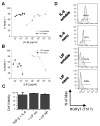
IL-6-nano induces RORγT expression following activation in the presence of TGF-β while LIF-nano suppresses IL-6-driven RORγT expression. (A) Naïve CD4+ T cells were stimulated for 72 hours with plate-bound anti-CD3, soluble anti-CD28, soluble IL-2, soluble TGF-β, and increasing doses of IL-6 or IL-6-nano. Expression of RORγT was assessed via flow cytometry and is shown on the y-axis. Nanoparticle dose of IL-6 was estimated to be approximately 2000 pg IL-6 per mg NP from (Fig. 2F). (B) Naïve CD4+ T cells were stimulated in same conditions as in (A) with the addition of 20 ng/ml IL-6 to the media. Nanoparticle dose of LIF was estimated to be 1000 pg LIF per mg NP from (Fig. 2E). (C) Cell viability at maximum shown doses was assessed by MTT assay. (D) Representative FACS plots of maximally effective doses from (A) and (B) demonstrate opposing effects of LIF and IL-6 on RORγT expression.
Figure 5
Figure 5. CD4-targeted NPs bind to CD4+ T cells
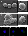
CD4+ T cells were imaged by SEM and fluorescent microscopy stimulation after incubation with or without CD4-targeted NPs. (A) Naïve CD4+ T cells were stimulated for 24 hours in the presence of antibodies only then fixed in 4% paraformaldehyde + 0.1% glutaraldehyde and imaged via SEM. (B) SEM of CD4+ T cells stimulated in the presence of CD4-targeted LIF-nano. For fluorescent experiments, CD4+ T cells were incubated alone (C) or with CD4-targeted nanoparticles encapsulating coumarin 6 (green) for 2 hours at either (D) 37°C or (E) 4°C. Cells were fixed with 4% paraformaldehyde and permeabilized using 0.1% Triton-X100. Cell nuclei and cytoskeleton were stained using DAPI (blue) and Texas-Red phalloidin (red), and cells were imaged at 40× magnification.
Figure 6
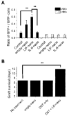
Figure 6. Localized delivery of LIF from CD4-targeted nanoparticles supports in vivo expansion of Foxp3+ T cells and supports allograft survival
(A) DBA/2 splenocytes were incubated for 15 minutes with CD4-targeted empty-nano, or LIF-nano, then infused (107 cells/mouse, i.v.) into BALB/c Foxp3-GFP mice (n=3 per group). Host lymph node cells were harvested 5 days later, and ratios of GFP+ vs. GFP- cells were calculated by FACS in the donor specific Vβ6 (black fill) or Vβ8 (striped fill) CD4+ T cell compartments (mean ± s.d.). Statistical significance was calculated by two-tailed t-test (** p < 0.01). (B) Vascularised heart grafts from BALB/c to CBA mice were rejected at 7d (no treatment controls; n=29: mean survival 6.86d). Animals received DST in similar fashion to the experiment in (A). A single i.v. dose of empty LIF-nano targeted to CD4 (empty nano; n=3: mean survival 7d) at time of grafting had no effect of graft survival. Donor-specific transfusion (DST only) alone had no effect as well (n=2: mean survival 7d). DST combined with LIF-nano therapy (DST + LIF-nano; n=3: mean survival 12.6d) resulted in significant prolongation of graft survival relative to the untreated control group (p=<0.01): analyses include Pearson Chi-Square p<0.01; and pExact statistics either one- or two-tailed p<0.01.
Figure 7
Figure 7. LIF-nano induce expansion of FOXP3+ CD4+ CD25+ T lymphocytes of the rhesus monkey
Peripheral blood lymphocytes (pbl) from 2 alloreactive rhesus monkeys were set up in a one way MLR (see methods). After 7d the cultures were boosted with irradiated donor pbl. Nine culture conditions were applied, as indicated. Both the LIF-nano and the empty-nano were targeted to CD4. The culture sets of 9 conditions were replicated for assaying by flow cytometry at 7d, prior to boosting (left-hand panels), and at 11d, 4d after boost (right-hand panels). (A) Analysis of the CD4+ cell population for FOXP3 (axis) and CD25 (abcissa) expression at 7d, and at 11d. (B) Total numbers of CD4+ cells in each culture at 11d shown as (i) all CD4+ cells (white fill); (ii) all dual CD4+ FOXP3+ cells (striped fill); and (iii) all triple positive CD4+ FOXP3+ CD25+ cells (black fill). Percentages are relative to controls.

References

    1. Riley JL, June CH, Blazar BR. Human T regulatory cell therapy: take a billion or so and call me in the morning. Immunity. 2009;30(5):656–65. - PMC - PubMed
    1. Masteller EL, Tang Q, Bluestone JA. Antigen-specific regulatory T cells--ex vivo expansion and therapeutic potential. Semin Immunol. 2006;18(2):103–10. - PubMed
    1. Zhou L, Chong MM, Littman DR. Plasticity of CD4+ T cell lineage differentiation. Immunity. 2009;30(5):646–55. - PubMed
    1. Littman DR, Rudensky AY. Th17 and regulatory T cells in mediating and restraining inflammation. Cell. 140(6):845–58. - PubMed
    1. Heinrich PC, et al. Principles of interleukin (IL)-6-type cytokine signalling and its regulation. Biochem J. 2003;374(Pt 1):1–20. - PMC - PubMed

Publication types

MeSH terms