

Signaling by FGFR2b controls the regenerative capacity of adult mouse incisors
- PMID: 20978072
- PMCID: PMC3049274
- DOI: 10.1242/dev.051672
Signaling by FGFR2b controls the regenerative capacity of adult mouse incisors
Abstract
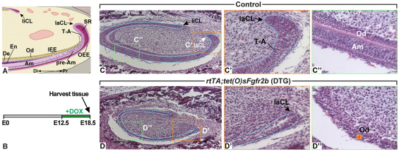
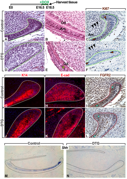
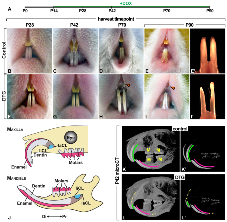
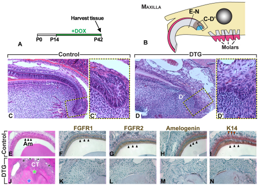
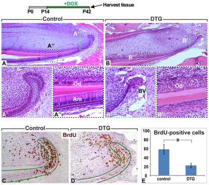
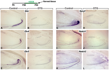
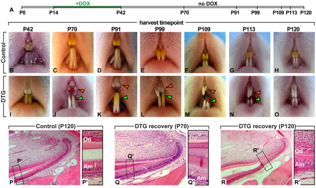
Rodent incisors regenerate throughout the lifetime of the animal owing to the presence of epithelial and mesenchymal stem cells in the proximal region of the tooth. Enamel, the hardest component of the tooth, is continuously deposited by stem cell-derived ameloblasts exclusively on the labial, or outer, surface of the tooth. The epithelial stem cells that are the ameloblast progenitors reside in structures called cervical loops at the base of the incisors. Previous studies have suggested that FGF10, acting mainly through fibroblast growth factor receptor 2b (FGFR2b), is crucial for development of the epithelial stem cell population in mouse incisors. To explore the role of FGFR2b signaling during development and adult life, we used an rtTA transactivator/tetracycline promoter approach that allows inducible and reversible attenuation of FGFR2b signaling. Downregulation of FGFR2b signaling during embryonic stages led to abnormal development of the labial cervical loop and of the inner enamel epithelial layer. In addition, postnatal attenuation of signaling resulted in impaired incisor growth, characterized by failure of enamel formation and degradation of the incisors. At a cellular level, these changes were accompanied by decreased proliferation of the transit-amplifying cells that are progenitors of the ameloblasts. Upon release of the signaling blockade, the incisors resumed growth and reformed an enamel layer, demonstrating that survival of the stem cells was not compromised by transient postnatal attenuation of FGFR2b signaling. Taken together, our results demonstrate that FGFR2b signaling regulates both the establishment of the incisor stem cell niches in the embryo and the regenerative capacity of incisors in the adult.
Figures
Similar articles
-
Bcl11b transcription factor plays a role in the maintenance of the ameloblast-progenitors in mouse adult maxillary incisors.Mech Dev. 2013 Sep-Oct;130(9-10):482-92. doi: 10.1016/j.mod.2013.05.002. Epub 2013 May 30. Mech Dev. 2013. PMID: 23727454
-
FGFR2 in the dental epithelium is essential for development and maintenance of the maxillary cervical loop, a stem cell niche in mouse incisors.Dev Dyn. 2009 Feb;238(2):324-30. doi: 10.1002/dvdy.21778. Dev Dyn. 2009. PMID: 18985768 Free PMC article.
-
Terminal end bud maintenance in mammary gland is dependent upon FGFR2b signaling.Dev Biol. 2008 May 1;317(1):121-31. doi: 10.1016/j.ydbio.2008.02.014. Epub 2008 Feb 21. Dev Biol. 2008. PMID: 18381212
-
FGF10: A multifunctional mesenchymal-epithelial signaling growth factor in development, health, and disease.Cytokine Growth Factor Rev. 2016 Apr;28:63-9. doi: 10.1016/j.cytogfr.2015.10.001. Epub 2015 Oct 31. Cytokine Growth Factor Rev. 2016. PMID: 26559461 Review.
-
Fibroblast growth factor signaling in mammalian tooth development.Odontology. 2014 Jan;102(1):1-13. doi: 10.1007/s10266-013-0142-1. Epub 2013 Dec 17. Odontology. 2014. PMID: 24343791 Free PMC article. Review.
Cited by
-
Fibroblast growth factor 10 alters the balance between goblet and Paneth cells in the adult mouse small intestine.Am J Physiol Gastrointest Liver Physiol. 2015 Apr 15;308(8):G678-90. doi: 10.1152/ajpgi.00158.2014. Epub 2015 Feb 26. Am J Physiol Gastrointest Liver Physiol. 2015. PMID: 25721301 Free PMC article.
-
Fgf10/Fgfr2b Signaling in Mammary Gland Development, Homeostasis, and Cancer.Front Cell Dev Biol. 2020 Jun 26;8:415. doi: 10.3389/fcell.2020.00415. eCollection 2020. Front Cell Dev Biol. 2020. PMID: 32676501 Free PMC article. Review.
-
Sox2+ stem cells contribute to all epithelial lineages of the tooth via Sfrp5+ progenitors.Dev Cell. 2012 Aug 14;23(2):317-28. doi: 10.1016/j.devcel.2012.05.012. Epub 2012 Jul 19. Dev Cell. 2012. PMID: 22819339 Free PMC article.
-
On the cutting edge of organ renewal: Identification, regulation, and evolution of incisor stem cells.Genesis. 2014 Feb;52(2):79-92. doi: 10.1002/dvg.22732. Epub 2013 Dec 14. Genesis. 2014. PMID: 24307456 Free PMC article. Review.
-
Evidence for the involvement of fibroblast growth factor 10 in lipofibroblast formation during embryonic lung development.Development. 2015 Dec 1;142(23):4139-50. doi: 10.1242/dev.109173. Epub 2015 Oct 28. Development. 2015. PMID: 26511927 Free PMC article.
References
-
- Bitgood M. J., McMahon A. P. (1995). Hedgehog and Bmp genes are coexpressed at many diverse sites of cell-cell interaction in the mouse embryo. Dev. Biol. 172, 126-138 - PubMed
-
- de Maximy A. A., Nakatake Y., Moncada S., Itoh N., Thiery J. P., Bellusci S. (1999). Cloning and expression pattern of a mouse homologue of drosophila sprouty in the mouse embryo. Mech. Dev. 81, 213-216 - PubMed
-
- De Moerlooze L., Spencer-Dene B., Revest J., Hajihosseini M., Rosewell I., Dickson C. (2000). An important role for the IIIb isoform of fibroblast growth factor receptor 2 (FGFR2) in mesenchymal-epithelial signalling during mouse organogenesis. Development 127, 483-492 - PubMed
Publication types
MeSH terms
Substances
Grants and funding
LinkOut - more resources
Full Text Sources
Molecular Biology Databases
