. 2010 Nov;137(22):3795-800.
doi: 10.1242/dev.053447.

Hox11 genes establish synovial joint organization and phylogenetic characteristics in developing mouse zeugopod skeletal elements

Affiliations

Hox11 genes establish synovial joint organization and phylogenetic characteristics in developing mouse zeugopod skeletal elements

Eiki Koyama et al. Development. 2010 Nov.

Abstract

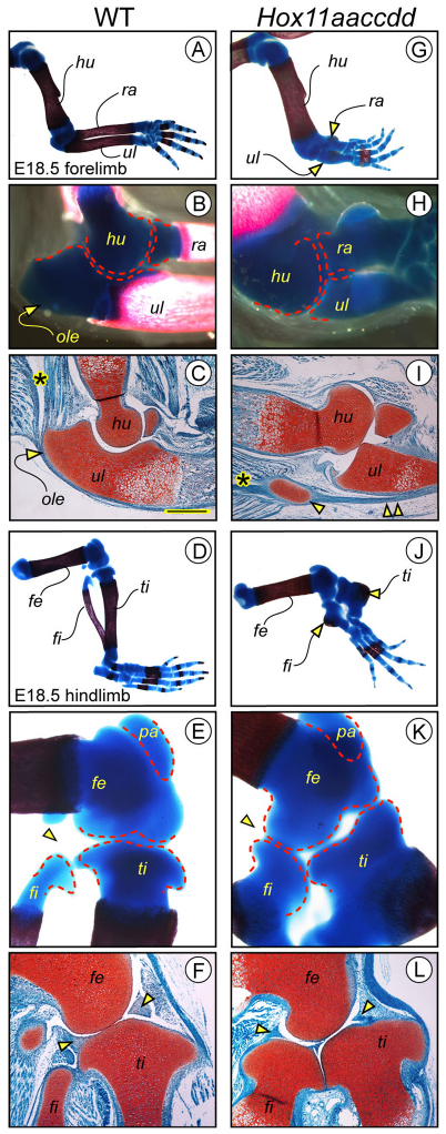
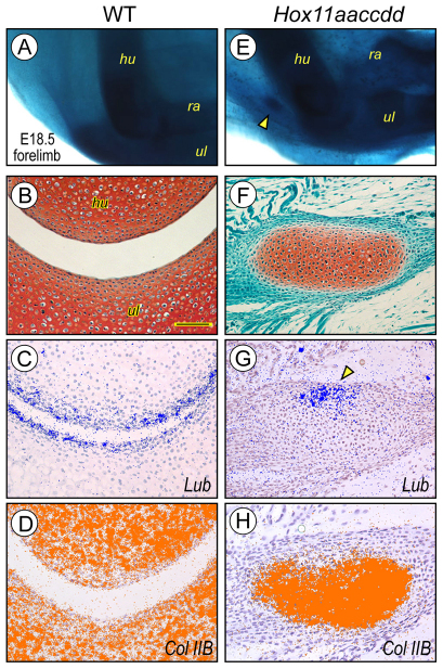
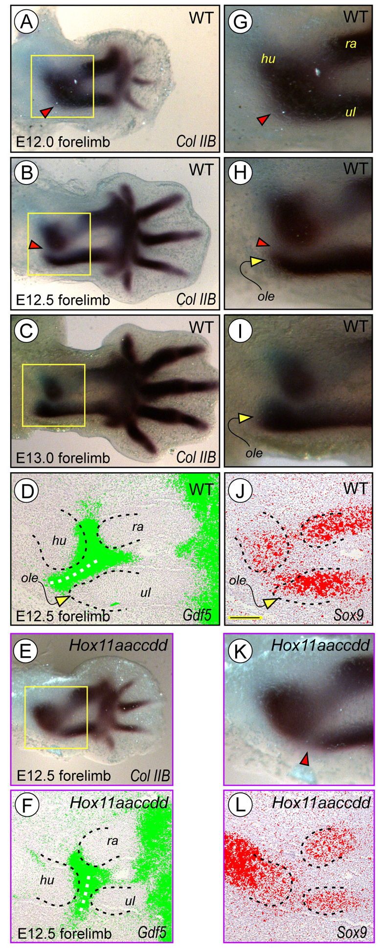
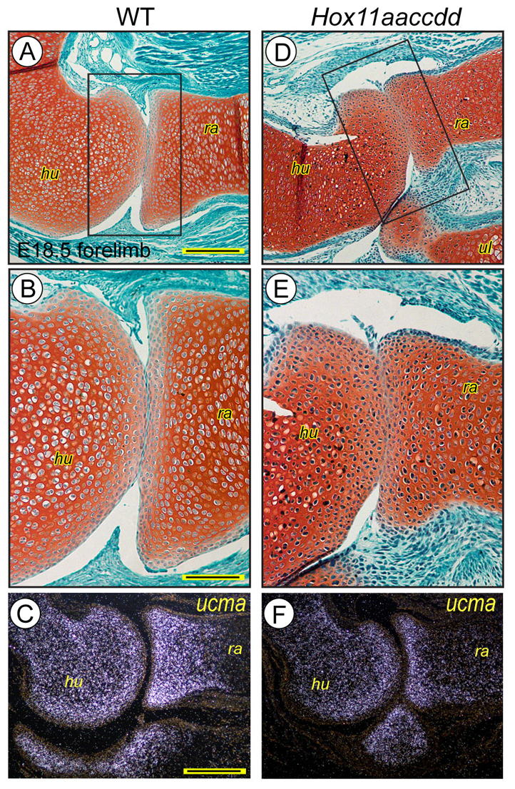
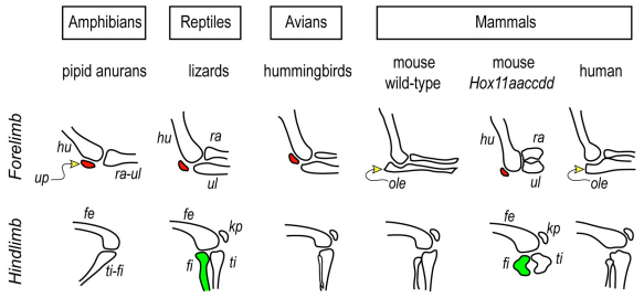
Hox11 genes are essential for zeugopod skeletal element development but their roles in synovial joint formation remain largely unknown. Here, we show that the elbow and knee joints of mouse embryos lacking all Hox11 paralogous genes are specifically remodeled and reorganized. The proximal ends of developing mutant ulna and radius elements became morphologically similar and formed an anatomically distinct elbow joint. The mutant ulna lacked the olecranon that normally attaches to the triceps brachii muscle tendon and connects the humerus to the ulna. In its place, an ulnar patella-like element developed that expressed lubricin on its ventral side facing the joint and was connected to the triceps muscle tendon. In mutant knees, both tibia and fibula fully articulated with an enlarged femoral epiphyseal end that accommodated both elements, and the neo-tripartite knee joint was enclosed in a single synovial cavity and displayed an additional anterior ligament. The mutant joints also exhibited a different organization of the superficial zone of articular cartilage that normally exerts an anti-friction function. In conclusion, Hox11 genes co-regulate and coordinate the development of zeugopod skeletal elements and adjacent elbow and knee joints, and dictate joint identity, morphogenesis and anatomical and functional organization. Notably, the ulnar patella and tripartite knee joints in the mouse mutants actually characterize several lower vertebrates, including certain reptiles and amphibians. The re-emergence of such anatomical structures suggests that their genetic blueprint is still present in the mouse genome but is normally modified to the needs of the mammalian joint-formation program by distinct Hox11 function.


Figures

Fig. 1.
Fig. 1.
Elbow and knee joints are remodeled and reorganized in triple Hox11 mutants. (A-F) E18.5 wild-type forelimbs and hindlimbs were stained with Alizarin Red and Alcian Blue to reveal humerus (hu), radius (ra), ulna (ul), femur (fe), tibia (ti) and fibula (fi) elements and photographed at low (A,D) and high (B,E) magnification. Companion specimens were processed for histochemical analysis of elbow (C) and knee (F) joints by Safranin-O fast green staining. Note the prominent olecranon (ole) attached to the triceps brachii muscle tendon (C, asterisk) and surrounding the humeral epiphysis, the fibula articulating with the lateral condyle of the tibia (E, arrowhead), and the bipartite knee joint contained within its synovial cavity (F, opposing yellow arrowheads). (G-L) Limbs from Hox11aaccdd mutant littermates exhibit hypomorphic and wholly cartilaginous radius and ulna (G,H) and tibia and fibula (J,K) elements. Note that the olecranon is absent and replaced by the ectopic ulnar patella (I, arrowhead), attached by the triceps brachii muscle tendon (I, asterisk) connecting the humerus to the ulna, and that the tibia and enlarged fibula fully articulate with a remodeled and enlarged distal femoral epiphysis (K, arrowhead) and form a neo-tripartite joint contained within a single synovial cavity (L, opposing yellow arrowheads). pa, patella. Scale bar: 250 μm in C,F,I,L.
Fig. 2.
Fig. 2.
The ectopic ulnar patella has phenotypic characteristics of a functional articular element. Whole-mount embryos and serial elbow region sections from E18.5 wild-type embryos (A-D) and Hox11aaccdd mutants (E-H) were examined by Alcian Blue staining (A,E), Safranin-O fast green staining (B,F) or in situ hybridization (C,D,G,H). Positive hybridization signal is presented in computer-generated coloring. The wild-type elbow joint and tissues exhibit stereotypic organization (A,B) and strong expression of lubricin (Lub; blue) and collagen IIB (Col IIB; orange) (C,D). The mutant elbow exhibits a distinct and sesamoid-like ulnar patella (E, arrowhead) never seen in wild types (A) that strongly expresses lubricin in its ventral side facing the elbow joint (G, arrowhead) and collagen IIB throughout its cartilaginous tissue (H). Scale bar: 100 μm in B-D,F-H.
Fig. 3.
Fig. 3.
The olecranon primordium is not specified in Hox11 mutants. E12.0, E12.5 and E13.0 wild-type (A-D,G-J) and triple-Hox11-null (E,F,K,L) forelimbs and sections thereof were examined by in situ hybridization. Note in E12.0 wild types that the collagen IIB-expressing skeletal elements are still fused and a prospective elbow joint and an interzone are undetectable (A,G, arrowheads). A Gdf5-expressing interzone and a collagen IIB- and Sox9-expressing olecranon (ole) primordium become appreciable by E12.5 (B,D,H,J, arrowheads and arrows, respectively) and are fully evident by E13.0 (C,I). In E12.5 mutants, however, a Gdf5-expressing interzone is present in between the collagen IIB-expressing elements but an olecranon primordium is not detectable (E,F,K,L). Note also the different orientation of the Gdf5-expressing interzone in wild-type and mutant joints (D,F, white dashed line). Positive hybridization signal is in green for Gdf5, red for Sox9 and brown/black for collagen IIB. Scale bar: 200 μm in D-F,J-L.
Fig. 4.
Fig. 4.
The articular superficial zone is sub-standard in Hox11 mutants. Sections of E18.5 wild-type (A-C) and Hox11-null (D-F) elbows were stained with Safranin-O fast green (A,B,D,E) or processed for Ucma expression analysis (C,F). Note that the superficial zone in mutants is ill-defined and does not exhibit differential matrix staining. In addition, the mutant epiphyseal portion displays reduced Ucma expression. Scale bars: 250 μm in A,D; 120 μm in B,E; 300 μm in C,F.
Fig. 5.
Fig. 5.
Schematic of possible phylogenetic links between wild-type and mutant joints. This schematic depicts the general organization and morphologies of elbow and knee joints in representative animal groups and species (Barnett and Lewis, 1958; Drapeau, 2004; Dye, 1987; Fujiwara et al., 2010; Haines, 1969; Soren and Waugh, 1994) compared with the joints in triple-Hox11-null mouse mutants. Most mammals have a conspicuous olecranon that encircles the distal end of the humerus, whereas the indicated groups of amphibians, reptiles and avians have a markedly smaller or even absent olecranon but have an ulnar patella (up, red). Thus, the presence of an ulnar patella and the absence of a clear olecranon in the Hox11 mutants suggest that their elbow joints have acquired characteristics of more-ancestral vertebrate groups. Absence of the olecranon and presence of an ulnar patella in certain species led to the suggestion that evolution of the olecranon might have involved fusion of the ulnar patella (a sesamoid) to the proximal end of the ulna (Barnett and Lewis, 1958), but this remains unclear (Haines, 1969). With regard to hind limbs, the tripartite knee joint in which fibula and tibia fully articulate with the femur within a single synovial cavity is characteristic of many reptiles and certain amphibians. Thus, the mutant mouse knee joint shares characteristics with those species. Note that the above scheme is not meant to be comprehensive and is used here to provide a phylogenetic representation and interpretation of the results. hu, humerus; ra, radius; ul, ulna; fe, femur; fi, fibula; ti, tibia; ole, olecranon; up, ulnar patella.

Similar articles

Cited by

References

    1. Archer C. W., Dowthwaite G. P., Francis-West P. (2003). Development of synovial joints. Birth Defects Research, Pt. C 69, 144-155 - PubMed
    1. Barnett C. H., Lewis O. J. (1958). The evolution of some traction epiphyses in birds and mammals. J. Anat. 92, 593-601 - PMC - PubMed
    1. Boulet A. M., Capecchi M. R. (2003). Multiple roles of Hoxa11 and Hoxd11 in the formation of the mammalian forelimb zeugopod. Development 131, 299-309 - PubMed
    1. Carroll S. B. (2008). Evo-Devo and an expanding evolutionary synthesis; a genetic theory of morphological evolution. Cell 134, 25-36 - PubMed
    1. Choocheep K., Hatano S., Takagi H., Watanabe H., Kimata K., Kongtawelert P., Watanabe H. (2010). Versican facilitates chondrocyte differentiation and regulates joint morphogenesis. J. Biol. Chem. 285, 21114-21125 - PMC - PubMed

Publication types