. 2010 Nov;3(11):1409-16.
doi: 10.1158/1940-6207.CAPR-10-0118. Epub 2010 Oct 26.

An MLH1 mutation links BACH1/FANCJ to colon cancer, signaling, and insight toward directed therapy

Affiliations

An MLH1 mutation links BACH1/FANCJ to colon cancer, signaling, and insight toward directed therapy

Jenny Xie et al. Cancer Prev Res (Phila). 2010 Nov.

Abstract

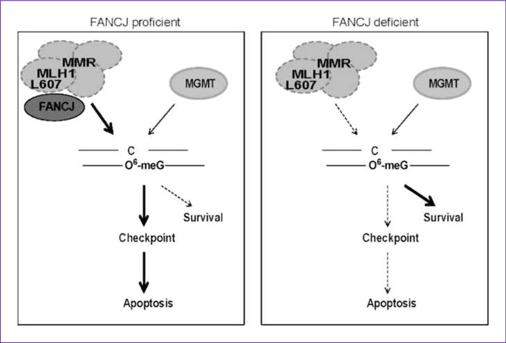
Defects in MLH1, as with other mismatch repair (MMR) proteins, are the primary cause of hereditary nonpolyposis colon cancer (HNPCC). Mutations in MMR genes often disrupt mismatch repair and MMR signaling functions. However, some HNPCC-associated mutations have unknown pathogenicity. Here, we uncover an MLH1 clinical mutation with a leucine (L)-to-histidine (H) amino acid change at position 607 that ablates MLH1 binding to FANCJ. Given that a DNA helicase is not essential for mammalian MMR in vitro, we considered that loss of MLH1 binding to FANCJ could alter MMR signaling. Consistent with this hypothesis, FANCJ-deficient cells exhibit delayed MMR signaling and apoptotic responses that generate resistance to agents that induce O(6)-methylguanine lesions. Our data indicate that the delay in MMR signaling provides time for the methylguanine methyltransferase (MGMT) enzyme to reverse DNA methylation. In essence, FANCJ deficiency alters the competition between two pathways: MGMT-prosurvival versus MMR-prodeath. This outcome could explain the HNPCC familial cancers that present as microsatellite stable and with intact MMR, such as MLH(L607H). Importantly, the link between FANCJ and HNPCC provides insight toward directed therapies because loss of the FANCJ/MLH1 interaction also uniquely sensitizes cells to DNA cross-linking agents.


Conflict of interest statement

Disclosure of Potential Conflicts of Interest

No potential conflicts of interest were disclosed.

Figures

Fig. 1
Fig. 1
MLH1 L607H mutant is defective for FANCJ binding and alters DNA damage response. A, MLH1 cartoon showing the FANCJ and PMS2 interacting domains. 293T cells (B) or HCT116 cells (C) cotransfected with PMS2 with different versions of MLH1 were collected and analyzed by Western blot with the indicated antibodies or immunoprecipitated with MLH1 or FANCJ antibody and blotted with the indicated antibodies. Transfected HCT116 cells were plated at low density, treated with the indicated doses of (D) MMC or (E) MNU with MGMT active or inhibited, and allowed to grow for 5 d. Percent growth was analyzed by collecting and counting the cells or by measuring ATP content.
Fig. 2
Fig. 2
When MGMT is active, FA-J cells are uniquely resistant to MNU. A, FANCJ-null FA-J (EUFA30F) cells were complemented with vector or FANCJWT; cells were either collected and analyzed by immunoblot with the indicated antibodies or (B) plated at low density, treated with the indicated doses of MNU with MGMT active or inhibited, and allowed to grow for 8 d. The cells were collected and counted to analyze percent growth. C, FA-D2 (PD20) and FA-D2+FANCD2 cells were either collected and analyzed by immunoblot with the indicated antibodies or treated with the indicated dose of MNU. The cells were collected and counted to analyze percent growth 8d later. D, Fibroblast (2822), FA-J (2833), FA-J (EUFA30F), FA-C (PD331), or FA-G (PD352) cells were plated at low density, treated with the indicated dose of MNU; and allowed to grow for 8 d. Percent growth was assessed as described in B.
Fig. 3
Fig. 3
The helicase activity and interaction with MLH1 of FANCJ are required for its role in MNU response. A, FANCJ-null FA-J (EUFA30F) cells complemented with vector or FANCJWT were treated with 1 mmol/L MNU with MGMT active. Cells were collected at various time points and analyzed by immunoblot with the indicated antibodies. B, vector or FANCJWT complemented FA-J cells were plated at low density and treated with the indicated dose of N-(2-chloroethyl)-N′-cyclohexyl-N-nitrosourea (CCNU) or N-methyl-N-nitro-N-nitrosoguanidine (MNNG). Cells were collected and counted to analyze percent growth 4 to 8 d later. C, FA-J cells were complemented with vector, FANCJWT, FANCJK52R, or FANCJK141/142A cells, which were either collected and analyzed by immunoblot with the indicated antibodies or (D) plated at low density and treated with the indicated dose of MNU. Percent growth was assessed as described in B.
Fig. 4
Fig. 4
FANCJ functions with MMR proteins in MNU response. A, MCF7 cells stably expressing shRNA reagents targeting control, MLH1, or FANCJ were either analyzed by immunoblot with the indicated antibodies or B, apoptosis was analyzed after cells were treated with the indicated dose of MNU with MGMT active or inhibited and allowed to grow for the indicated time periods. The cells were collected, fixed, and stained with Annexin V and propidium iodide (percent apoptotic cells were analyzed by fluorescence-activated cell sorting). C and D, stable MCF7 cells were treated with the indicated dose of MNU with MGMT active or inhibited. Cells were fixed and stained with Giemsa 8 d later, and then the colonies were counted. The graph represents three independent experiments. E, stable MCF7 cells were treated with the indicated dose of hydroxyurea (HU) or MNU, collected after 24 h, and analyzed by immunoblot with the indicated antibodies. F, the graph represents the densitometry of pChk1 versus total Chk1.
Fig. 5
Fig. 5
Model showing the enhancement of MMR signaling and apoptosis by FANCJ. In FANCJ-proficient cells, MMR proteins efficiently process O6-meG lesions leading to checkpoint activation and apoptosis. In FANCJ-deficient cells, MMR proteins are less efficient at processing O6-meG lesions, allowing more time for MGMT processing and thus enhancing cell survival. This study identified MLH1 L607 as a residue required for MLH1 binding to FANCJ.

Comment in

References

    1. O’Brien V, Brown R. Signalling cell cycle arrest and cell death through the MMR System. Carcinogenesis. 2006;27:682–92. - PubMed
    1. Kat A, Thilly WG, Fang WH, Longley MJ, Li GM, Modrich P. An alkylation-tolerant, mutator human cell line is deficient in strand-specific mismatch repair. Proc Natl Acad Sci U S A. 1993;90:6424–8. - PMC - PubMed
    1. Branch P, Aquilina G, Bignami M, Karran P. Defective mismatch binding and a mutator phenotype in cells tolerant to DNA damage. Nature. 1993;362:652–4. - PubMed
    1. Schofield MJ, Hsieh P. DNA mismatch repair: molecular mechanisms and biological function. Annu Rev Microbiol. 2003;57:579–608. - PubMed
    1. Karran P, Marinus MG. Mismatch correction at O6-methylguanine residues in E. coli DNA. Nature. 1982;296:868–9. - PubMed

Publication types

MeSH terms

Substances