. 2010 Nov;3(11):1462-72.
doi: 10.1158/1940-6207.CAPR-10-0037. Epub 2010 Oct 26.

Thiocolchicoside exhibits anticancer effects through downregulation of NF-κB pathway and its regulated gene products linked to inflammation and cancer

Affiliations

Thiocolchicoside exhibits anticancer effects through downregulation of NF-κB pathway and its regulated gene products linked to inflammation and cancer

Simone Reuter et al. Cancer Prev Res (Phila). 2010 Nov.

Abstract

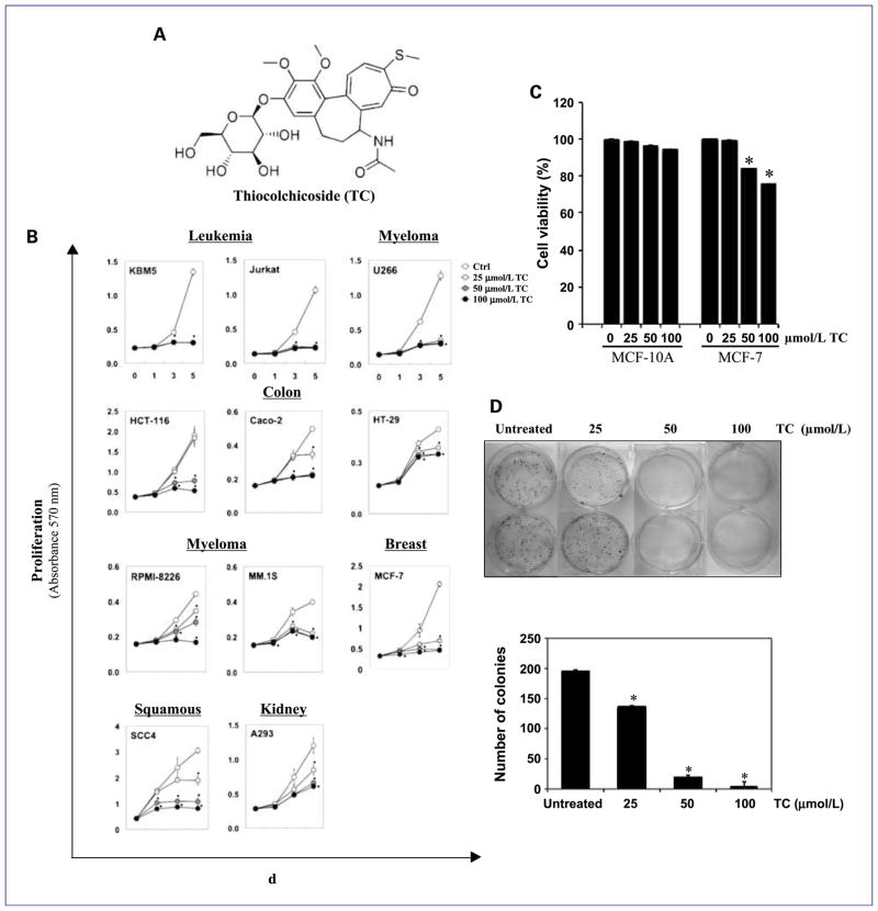
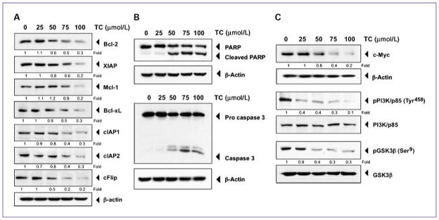
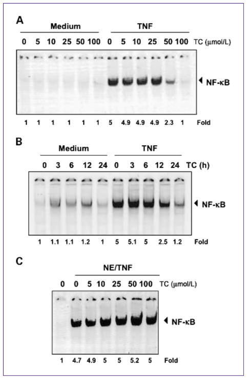
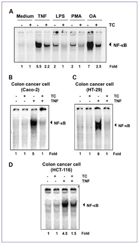
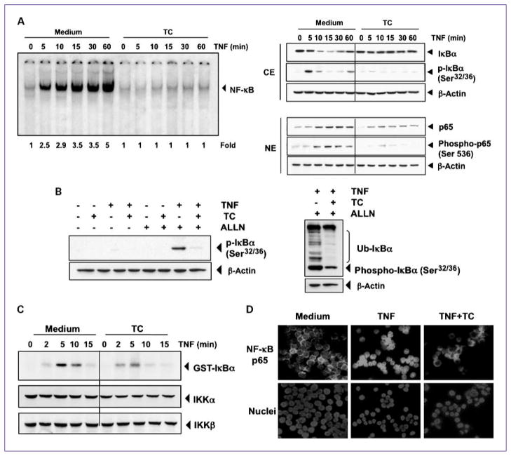
The discovery of new uses for older, clinically approved drugs is one way to expedite drug development for cancer. Thiocolchicoside, a semisynthetic colchicoside from the plant Gloriosa superba, is a muscle relaxant and used to treat rheumatologic and orthopedic disorders because of its analgesic and anti-inflammatory mechanisms. Given that activation of the transcription factor NF-κB plays a major role in inflammation and tumorigenesis, we postulated that thiocolchicoside would inhibit NF-κB and exhibit anticancer effects through the modulation of NF-κB-regulated proteins. We show that thiocolchicoside inhibited proliferation of leukemia, myeloma, squamous cell carcinoma, breast, colon, and kidney cancer cells. Formation of tumor colonies was also suppressed by thiocolchicoside. The colchicoside induced apoptosis, as indicated by caspase-3 and poly(ADP-ribose) polymerase cleavage, and suppressed the expression of cell survival [e.g., Bcl-2, X-linked inhibitor of apoptosis (XIAP), MCL-1, bcl-xL, cIAP-1, cIAP-2, and cFLIP] proteins. Cell proliferation biomarkers such as c-MYC and phosphorylation of phosphoinositide 3-kinase and glycogen synthase kinase 3β were also blocked by thiocolchicoside. Because most cell survival and proliferation gene products are regulated by NF-κB, we studied the effect of thiocolchicoside on this transcription factor and found that thiocolchicoside inhibited NF-κB activation, degradation of inhibitory κBα (IκBα), IκBα ubiquitination, and phosphorylation, abolished the activation of IκBα kinase, and suppressed p65 nuclear translocation. This effect of thiocolchicoside on the NF-κB pathway led to inhibition of NF-κB reporter activity and cyclooxygenase-2 promoter activity. Our results indicate that thiocolchicoside exhibits anticancer activity through inhibition of NF-κB and NF-κB-regulated gene products, which provides novel insight into a half-century old drug.


Conflict of interest statement

Disclosure of Potential Conflicts of Interest

No potential conflicts of interest were disclosed.

Figures

Fig. 1
Fig. 1
Thiocolchicoside suppresses cell proliferation and colony formation of various cancer cell lines. A, the structure of thiocolchicoside. B, thiocolchicoside inhibits cell proliferation of various cancer cell lines. Leukemia (KBM5, Jurkat), colon (HCT-116, Caco-2, HT-29), myeloma (U266, RPMI-8266, MM.1S), breast (MCF-7), squamous (SCC4), and kidney (A293) cells were treated with 25, 50, and 100 μmol/L thiocolchicoside for 1, 3, and 5 d, and cell proliferation was assessed by the MTT method. C, thiocolchicoside has no effect on normal cells. MCF-10A (nontransformed breast epithelial cells) cells and MCF-7 (transformed breast epithelial cells) cells were treated with 25, 50, and 100 μmol/L thiocolchicoside for 24 h, and cell viability was assessed by the MTT method. D, thiocolchicoside inhibits colony formation of HCT-116 cells. HCT-116 cells were plated in six-well plates and treated with 25, 50, and 100 μmol/L of thiocolchicoside for 24 h. After 1 d, medium was changed, and cells were incubated for 9 d for colony formation (top). After 9 d, cells were stained with crystal violet, and number of colonies was counted (bottom). Data are presented as mean (±SD), and * indicates P < 0.05, when compared with control.
Fig. 2
Fig. 2
Thiocolchicoside induces apoptosis and inhibits proteins involved in cell proliferation. A, thiocolchicoside downregulates the expression of antiapoptotic proteins. KBM5 cells were treated with 25, 50, 75, and 100 μmol/L of thiocolchicoside for 24 h; whole-cell extracts were prepared and analyzed by Western blot using antibodies against Bcl-2, XIAP, Mcl-1, Bcl-xL, cIAP1, cIAP2, and cFlip. B, thiocolchicoside induces apoptosis. KBM5 cells were treated with 25, 50, 75, and 100 μmol/L of thiocolchicoside for 24 h. Whole-cell extracts were prepared and analyzed by Western blot using antibodies against PARP and caspase-3. C, thiocolchicoside downregulates the expression of proteins involved in cell proliferation. KBM5 cells were treated with 25, 50, 75, and 100 μmol/L of thiocolchicoside for 24 h. Whole-cell extracts were prepared and analyzed by Western blot using antibodies against c-Myc, the phosphorylated PI3K/p85, and phosphorylated GSK3β proteins. Unphosphorylated proteins of PI3K/p85 and GSK3β as well as β-actin were used as a loading control. Numbers below each panel indicate fold differences after normalization to β-actin.
Fig. 3
Fig. 3
Thiocolchicoside inhibits TNF-dependent NF-κB activation. A, thiocolchicoside inhibits TNF-dependent NF-κB activation in a dose-dependent manner. KBM5 cells were preincubated with indicated concentrations of thiocolchicoside for 24 h, treated with 0.1 nmol/L TNF for 30 min, and then subjected to EMSA to test for NF-κB activation. B, thiocolchicoside inhibits TNF-dependent NF-κB activation in a time-dependent manner. KBM5 cells were preincubated with 100 μmol/L thiocolchicoside for the indicated times, treated with 0.1 nmol/L TNF for 30 min, and then subjected to EMSA to test for NF-κB activation. C, the direct effect of thiocolchicoside on NF-κB complex was investigated. Nuclear extracts were prepared from untreated cells or cells treated with 0.1 nmol/L TNF and incubated for 30 min with the indicated concentrations of thiocolchicoside. They were then assayed for NF-κB activation by EMSA. Numbers below panels indicate fold differences normalized to the control.
Fig. 4
Fig. 4
Thiocolchicoside-induced NF-κB inhibition is neither inducer nor cell type specific. A, thiocolchicoside suppresses NF-κB activation by different stimuli. KBM5 cells were preincubated with 100 μmol/L thiocolchicoside for 24 h and then treated with 0.1 nmol/L TNF or 10 μg/mL LPS for 30 min, 500 nmol/L OA for 4 h or 25 μg/mL PMA for 2 h. The cells were then analyzed for NF-κB activation by EMSA. B–D, thiocolchicoside induced NF-κB inhibition is not cell type specific. Caco-2, HT-29, and HCT-116 cells were incubated with 100 μmol/L thiocolchicoside for 24 h and then incubated with 0.1 nmol/L TNF for 30 min. Nuclear extracts were then prepared and assayed for NF-κB activation by EMSA. Numbers below panels indicate fold differences normalized to the control.
Fig. 5
Fig. 5
Thiocolchicoside inhibits TNF-dependent IκBα phosphorylation, IκBα degradation, p65 phosphorylation, and p65 nuclear translocation. A, thiocolchicoside inhibits TNF-induced activation of NF-κB. KBM-5 cells were incubated with 100 μmol/L thiocolchicoside for 24 h, treated with 0.1 nmol/L TNF for the indicated times, and then analyzed for NF-κB activation by EMSA (left). A, right, effect of thiocolchicoside on TNF-induced IκBα degradation, p65 phosphorylation, and p65 nuclear translocation. Cells were incubated with 100 μmol/L thiocolchicoside for 24 h and treated with 0.1 nmol/L TNF for the indicated times. Cytoplasmic extracts (CE) and nuclear extracts (NE) were prepared, fractionated on SDS-PAGE, and electrotransferred to nitrocellulose membrane. Western blot analysis was done using the indicated antibody. An anti-β-actin antibody was the loading control. B, effect of thiocolchicoside on the phosphorylation of IκBα by TNF. Cells were preincubated with 100 μmol/L thiocolchicoside for 24 h, incubated with 50 μg/mL ALLN for 30 min, and then treated with 0.1 nmol/L TNF for 10 min. Cytoplasmic extracts were fractionated and then subjected to Western blot analysis using a phospho-specific anti-IκBα antibody. An anti-β-actin antibody was used as loading control (left). B, right, thiocolchicoside inhibits ubiquitination of IκBα. Cells were preincubated with 100 μmol/L thiocolchicoside for 24 h, incubated with 50 μg/mL ALLN for 30 min, and then treated with 0.1 nmol/L TNF for 10 min. Cytoplasmic extracts were fractionated and then subjected to Western blot analysis using a phospho-specific anti-IκBα antibody. An anti-β-actin antibody used as the loading control. C, direct effect of thiocolchicoside on IKK activation induced by TNF. Whole-cell extracts were immunoprecipitated with antibody against IKKβ and analyzed by an immune complex kinase assay. To examine the effect of thiocolchicoside on the level of expression of IKK proteins, whole-cell extracts were fractionated on SDS-PAGE and examined by Western blot analysis using anti-IKKα and anti-IKKβ antibodies. D, immunocytochemical analysis of p65 localization. Cells were incubated with 100 μmol/L thiocolchicoside for 24 h and then treated with 1 nm TNF for 15 min. Cells were subjected to immunocytochemical analysis.
Fig. 6
Fig. 6
Thiocolchicoside represses NF-κB–dependent reporter gene expression induced by TNF and by overexpression of various signaling intermediates. A, thiocolchicoside inhibits the NF-κB–dependent reporter gene expression induced by TNF. A293 cells were transiently transfected with a NF-κB–containing plasmid for 24 h. After transfection, the cells were incubated with the indicated concentrations of thiocolchicoside for 24 h and then treated with 1 nmol/L TNF for an additional 24 h. The supernatants of the culture media were assayed for SEAP activity. Data are presented as mean (±SD). B, thiocolchicoside inhibits the NF-κB–dependent reporter gene expression induced by TNF, TNFR1, TRADD, TRAF2, NIK, IKK, and p65. Cells were transiently transfected with a NF-κB–containing plasmid alone or with the indicated plasmids. After transfection, cells were incubated with 100 μmol/L thiocolchicoside for 24 h and then incubated with the relevant plasmid for an additional 24 h. TNF-treated cells were incubated with 100 μmol/L thiocolchicoside for 24 h and then treated with 1 nmol/L TNF for an additional 24 h. The supernatants of the culture media were assayed for SEAP activity. Data are presented as mean (±SD). C, thiocolchicoside inhibits the COX-2 promoter activity induced by TNF. Cells were transiently transfected with a COX-2 promoter linked to the luciferase reporter gene plasmid for 24 h and treated with the indicated concentrations of thiocolchicoside for 24 h. Cells were then treated with 1 nmol/L TNF for an additional 24 h, lysed, and subjected to a luciferase assay. Data are presented as mean (±SD), and * indicates P < 0.05 when compared with their respective control.

References

    1. Janbroers JM. Review of the toxicology, pharmacodynamics and pharmacokinetics of thiocolchicoside, a GABA-agonist muscle relaxant with anti-inflammatory and analgesic actions. Acta Therapeutica. 1987;13:221–35.
    1. Tuzun F, Unalan H, Oner N, et al. Multicenter, randomized, double-blinded, placebo-controlled trial of thiocolchicoside in acute low back pain. Joint Bone Spine. 2003;70:356–61. - PubMed
    1. Ketenci A, Ozcan E, Karamursel S. Assessment of efficacy and psychomotor performances of thiocolchicoside and tizanidine in patients with acute low back pain. Int J Clin Pract. 2005;59:764–70. - PubMed
    1. Soonawalla DF, Joshi N. Efficacy of thiocolchicoside in Indian patients suffering from low back pain associated with muscle spasm. J Indian Med Assoc. 2008;106:331–5. - PubMed
    1. Balduini W, Cimino M, Depoortere H, Cattabeni F. Characterization of [3H]thiocolchicoside binding sites in rat spinal cord and cerebral cortex. Eur J Pharmacol. 1999;376:149–57. - PubMed

Publication types