. 2011 Jan;300(1):E103-10.
doi: 10.1152/ajpendo.00427.2010. Epub 2010 Oct 26.

APPL1 mediates adiponectin-stimulated p38 MAPK activation by scaffolding the TAK1-MKK3-p38 MAPK pathway

Affiliations

APPL1 mediates adiponectin-stimulated p38 MAPK activation by scaffolding the TAK1-MKK3-p38 MAPK pathway

Xiaoban Xin et al. Am J Physiol Endocrinol Metab. 2011 Jan.

Abstract

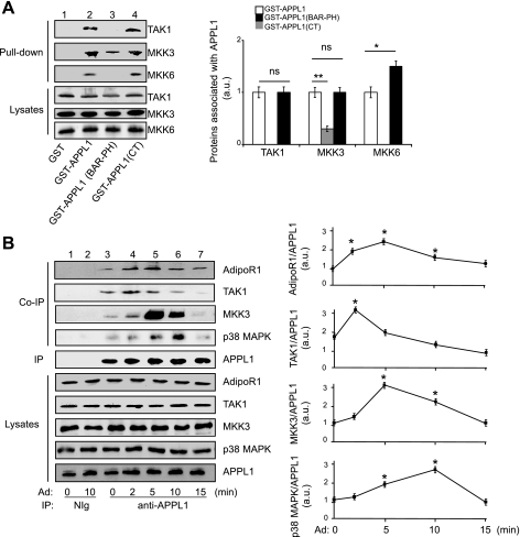
The adaptor protein APPL1 mediates the stimulatory effect of adiponectin on p38 mitogen-activated protein kinase (MAPK) signaling, yet the underlying mechanism remains unclear. Here we show that, in C(2)C(12) cells, overexpression or suppression of APPL1 enhanced or suppressed, respectively, adiponectin-stimulated p38 MAPK upstream kinase cascade, consisting of transforming growth factor-β-activated kinase 1 (TAK1) and mitogen-activated protein kinase kinase 3 (MKK3). In vitro affinity binding and coimmunoprecipitation experiments revealed that TAK1 and MKK3 bind to different regions of APPL1, suggesting that APPL1 functions as a scaffolding protein to facilitate adiponectin-stimulated p38 MAPK activation. Interestingly, suppressing APPL1 had no effect on TNFα-stimulated p38 MAPK phosphorylation in C(2)C(12) myotubes, indicating that the stimulatory effect of APPL1 on p38 MAPK activation is selective. Taken together, our study demonstrated that the TAK1-MKK3 cascade mediates adiponectin signaling and uncovers a scaffolding role of APPL1 in regulating the TAK1-MKK3-p38 MAPK pathway, specifically in response to adiponectin stimulation.


Figures

Fig. 1.
Fig. 1.
Transforming growth factor-β-activated kinase 1 (TAK1) and mitogen-activated protein kinase kinase 3 (MKK3) are essential in mediating adiponectin (Ad)-induced p38 mitogen-activated protein kinase (MAPK) activation. A: suppression of TAK1 or MKK3, but not MKK6, impaired Ad-induced p38 MAPK activation. C2C12 myocytes with TAK1, MKK3, or MKK6 suppressed by transfecting short hairpin RNA were serum starved for 6 h and treated with 1 μg/ml Ad for 10 min. The phosphorylated (P) (Thr180/Tyr182) and the protein levels of p38 MAPK, as well as TAK1, MKK3, MKK6, and tubulin, were detected by Western blot analysis with specific antibodies, as indicated. Graphic presentation indicated the effect of suppressing TAK1, MKK3, or MKK6 on the Ad-stimulated p38 MAPK activation shown in Western blot analysis. Values are means ± SE from three independent experiments. **P < 0.01. B: suppression of AMP-activated protein kinase (AMPK) expression does not affect Ad-stimulated p38 MAPK activation. The control or AMPKα2 RNA interference (RNAi) C2C12 myotubes were serum starved overnight and treated with 1 μg/ml Ad for 10 min. The P-p38 MAPK (Thr180/Tyr182) and the protein levels of p38 MAPK, as well as AMPKα2, were detected by Western blot analysis with specific antibodies, as indicated. Graphic presentation indicated the effect of suppressing AMPK on the Ad-stimulated p38 MAPK activation shown in Western blot analysis. Values are means ± SE from three independent experiments. **P < 0.01. C: Ad sequentially stimulates TAK1, MKK3, and p38 MAPK activities. C2C12 myotubes were serum starved overnight and treated with 1 μg/ml Ad for different times, as indicated. P-TAK1 (Thr184/187), P-p38 MAPK (Thr180/Tyr182), and their protein levels were detected by Western blot analysis with specific antibodies, as indicated. MKK3 was immunoprecipitated (IP) with an antibody specific to MKK3, and P-MKK3 was detected by Western blot with antibody to P-MKK3/6 (Ser189/207). Graphic presentation indicated the effect of Ad on activation of TAK1, MKK3, and p38 MAPK shown in Western blot analysis. Values are means ± SE from three independent experiments. The t-test was performed with the activity at each time point compared with the basal level. *P < 0.05 and **P < 0.01. Mock, mock control; Ctrl, control; ns, nonsignificant.
Fig. 2.
Fig. 2.
The role of APPL1 in mediating Ad-stimulated activation of TAK1-MKK3-p38 MAPK pathway. A: overexpression of APPL1 enhanced Ad-stimulated TAK1-MKK3-p38 MAPK cascade activation. C2C12 myocytes overexpressing pcDNA3.1/Myc-His(+)A vector (as a mock control) or pcDNA3.1/Myc-His(+)A/APPL1 were serum starved for 6 h and treated with 1 μg/ml Ad for different times, as indicated. P-TAK1 (Thr184/187) and P-p38 MAPK (Thr180/Tyr182) and their protein levels were detected by Western blot with specific antibodies, as indicated. MKK3 was IP with an antibody specific to MKK3, and P-MKK3 was detected by Western blot with antibody to P-MKK3/6 (Ser189/207). Graphic presentation indicated the effect of overexpression of APPL1 on Ad-stimulated activation of TAK1, MKK3, and p38 MAPK shown in the Western blot. Values are means ± SE from three independent experiments. The t-test was performed by comparing the activities between mock and overexpression group at each time point. *P < 0.05 and **P < 0.01. B: suppression of APPL1 expression impaired Ad-induced TAK1-MKK3-p38 MAPK cascade activation. The scrambled control or APPL1 RNAi C2C12 myotubes were serum starved overnight and treated with 1 μg/ml Ad for different times, as indicated. P-TAK1 (Thr184/187), P-p38 MAPK (Thr180/Tyr182), and their protein levels were detected by Western blot with specific antibodies, as indicated. MKK3 was IP with an antibody specific to MKK3, and P-MKK3 was detected by Western blot with antibody to P-MKK3/6 (Ser189/207). Graphic presentation indicated the effect of suppressing APPL1 expression on Ad-stimulated activation of TAK1, MKK3, and p38 MAPK shown in the Western blot. The t-test was performed by comparing the activities between the scramble and APPL1-RNAi group at each time point. Values are means ± SE from three independent experiments. *P < 0.05. au, Arbitrary units.
Fig. 3.
Fig. 3.
APPL1 is not involved in mediating TNFα-induced TAK1-MKK3-p38 MAPK activation. A: the scrambled control or APPL1 RNAi C2C12 myotubes were serum starved overnight and treated with TNFα (1 nM) for different times, as indicated. P-TAK1 (Thr184/187), P-p38 MAPK (Thr180/Tyr182), and their protein levels were detected by Western blot with specific antibodies, as indicated. MKK3 was IP with an antibody specific to MKK3, and P-MKK3 was detected by Western blot analysis with antibody to P-MKK3/6 (Ser189/207). Graphic presentation indicated the effect of suppressing APPL1 expression on TNFα-stimulated activation of TAK1, MKK3, and p38 MAPK shown in the Western blot. Values are means ± SE from three independent experiments. The t-test was performed by comparing the activities between scramble and RNAi groups at each time point. B: the effects of Ad and TNFα on the activation of α- and β-isoforms of p38 MAPK in C2C12 myotubes. C2C12 myotubes were serum starved overnight and treated with either Ad (1 μg/ml, 10 min) or TNFα (1 nM, 10 min). The α- and β-isoforms of p38 MAPK were IP, and P-p38 MAPK (Thr180/Tyr182) was detected by Western blot analysis with antibodies, as indicated. Graphic presentation indicated the effect of Ad or TNFα on activation of α- and β-isoforms of p38 MAPK shown in the Western blot. Values are means ± SE from three independent experiments. *P < 0.05. C: suppression of APPL1 impaired the effect of Ad on activation of the α- and β-isoform of p38 MAPK. The scrambled control or APPL1 RNAi C2C12 myotubes were serum starved overnight and treated with 1 μg/ml Ad for 10 min. The α- and β-isoforms of p38 MAPK were IP, and the P-p38 MAPK (Thr180/Tyr182) was detected by Western blot analysis with specific antibodies, as indicated. Graphic presentation indicates the effect of suppression of APPL1 on Ad-stimulated activation of the α- and β-isoforms of p38 MAPK shown in the Western blot. Values are means ± SE from three independent experiments. *P < 0.05. D: suppression of APPL1 had no effect of TNFα on activation of the β-isoform of p38 MAPK. The scrambled control or APPL1 RNAi C2C12 myotubes were serum starved overnight and treated with TNFα (1 nM) for 10 min. The β-isoform of p38 MAPK was IP, and the P-p38 MAPK (Thr180/Tyr182) was detected by Western blot analysis with specific antibodies, as indicated. Graphic presentation indicated the effect of suppression of APPL1 on TNFα-stimulated activation of β-isoform of p38 MAPK shown in the Western blot. Values are means ± SE from three independent experiments. *P < 0.05.
Fig. 4.
Fig. 4.
APPL1 acts as a scaffold protein by interacting with TAK1-MKK3-p38 MAPK. A: APPL1 interacts with TAK1, MKK3, and MKK6 in vitro. GST or GST-APPL1 (full-length, BAR-PH or CT truncations) fusion protein was incubated with cell lysates of C2C12 myotubes. Endogenous TAK1, MKK3, and MKK6 associated with recombinant GST-APPL1 (full-length or truncations) and their protein levels in the lysates were detected by Western blot analysis with specific antibodies, as indicated. Graphic presentation indicated the binding affinity between APPL1 fusion proteins with TAK1, MKK3, and MKK6 shown in the Western blot. Values are means ± SE from three independent experiments. *P < 0.05 and **P < 0.01. B: the effect of Ad on the interactions of APPL1 with Ad receptor 1 (AdipoR1), TAK1, MKK3, and p38 MAPK in cells. C2C12 myotubes were serum starved overnight and treated with 1 μg/ml Ad for the indicated time. Endogenous APPL1 was IP with the antibody specific to APPL1. Coimmunoprecipitated (Co-IP) AdipoR1, TAK1, MKK3, and p38 MAPK, as well as their protein levels in the cell lysates, were detected by Western blot analysis with specific antibodies, as indicated. Graphic presentation indicated the effect of Ad on the interaction of APPL1 with AdipoR1, TAK1, MKK3, and p38 MAPK shown in the Western blot. The t-test was performed by comparing the affinity with the basal conditions. Values are means ± SE from three independent experiments. *P < 0.05.
Fig. 5.
Fig. 5.
A model of APPL1-regulated TAK1-MKK3-p38 MAPK pathway in response to Ad stimulation. C', indicates the COOH terminus of AdipoR1. Under the basal condition, APPL1 binds with inactive TAK1 and weakly associates with MKK3 and p38 MAPK. On Ad stimulation, APPL1 interacts with AdipoR1, leading to activation of TAK1 and, subsequently, recruitment of MKK3 and p38 MAPK to form a complex consisting of AdipoR1, APPL1, TAK1, MKK3, and p38 MAPK. Once MKK3 is activated, TAK1 dissociates from APPL1, and the activity of TAK1 is rapidly downregulated. This process occurs concurrently with the dissociation of APPL1 complex from AdipoR1 and, in turn, stimulates the downstream events regulated by TAK1-MKK3-p38 MAPK pathway.

References

    1. Bae GU, Lee JR, Kim BG, Han JW, Leem YE, Lee HJ, Ho SM, Hahn MJ, Kang JS. Cdo interacts with APPL1 and activates AKT in myoblast differentiation. Mol Biol Cell 21: 2399–2411, 2010 - PMC - PubMed
    1. Beltowski J, Jamroz-Wiśniewska A, Widomska S. Adiponectin and its role in cardiovascular diseases. Cardiovasc Hematol Disord Drug Targets 8: 7–46, 2008 - PubMed
    1. Bhatnagar S, Kumar A, Makonchuk DY, Li H, Kumar A. Transforming growth factor-beta-activated kinase 1 is an essential regulator of myogenic differentiation. J Biol Chem 285: 6401–6411, 2010 - PMC - PubMed
    1. Chambers MA, Moylan JS, Smith JD, Goodyear LJ, Reid MB. Stretch-stimulated glucose uptake in skeletal muscle is mediated by reactive oxygen species and p38 MAP-kinase. J Physiol 587: 3363–3373, 2009 - PMC - PubMed
    1. Choo MK, Sakurai H, Koizumi K, Saiki I. TAK1-mediated stress signaling pathways are essential for TNF-alpha-promoted pulmonary metastasis of murine colon cancer cells. Int J Cancer 118: 2758–2764, 2006 - PubMed

Publication types

MeSH terms

LinkOut - more resources