. 2010 Nov;120(11):3953-68.
doi: 10.1172/JCI43490. Epub 2010 Oct 11.

Gene therapy using genetically modified lymphocytes targeting VEGFR-2 inhibits the growth of vascularized syngenic tumors in mice

Affiliations

Gene therapy using genetically modified lymphocytes targeting VEGFR-2 inhibits the growth of vascularized syngenic tumors in mice

Dhanalakshmi Chinnasamy et al. J Clin Invest. 2010 Nov.

Abstract

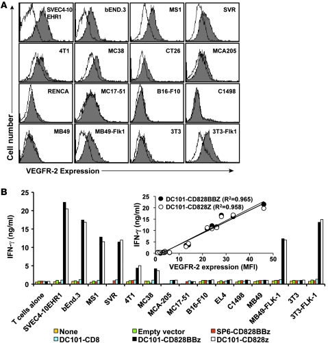
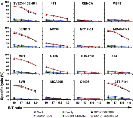
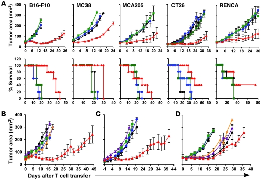
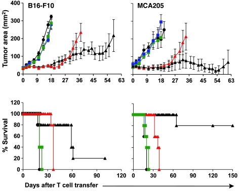
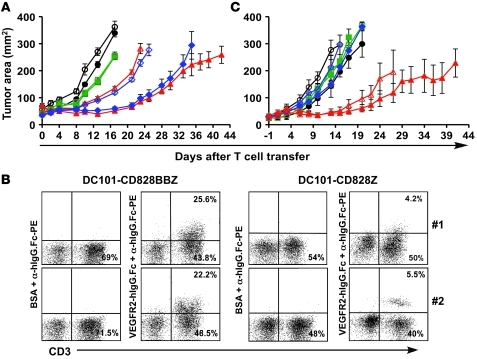
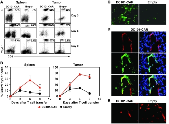
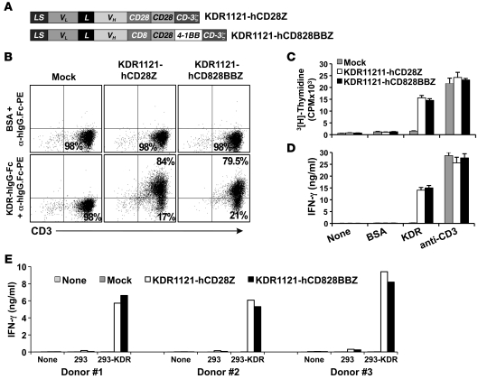
Immunotherapies based on adoptive cell transfer are highly effective in the treatment of metastatic melanoma, but the use of this approach in other cancer histologies has been hampered by the identification of appropriate target molecules. Immunologic approaches targeting tumor vasculature provide a means for the therapy of multiple solid tumor types. We developed a method to target tumor vasculature, using genetically redirected syngeneic or autologous T cells. Mouse and human T cells were engineered to express a chimeric antigen receptor (CAR) targeted against VEGFR-2, which is overexpressed in tumor vasculature and is responsible for VEGF-mediated tumor progression and metastasis. Mouse and human T cells expressing the relevant VEGFR-2 CARs mediated specific immune responses against VEGFR-2 protein as well as VEGFR-2-expressing cells in vitro. A single dose of VEGFR-2 CAR-engineered mouse T cells plus exogenous IL-2 significantly inhibited the growth of 5 different types of established, vascularized syngeneic tumors in 2 different strains of mice and prolonged the survival of mice. T cells transduced with VEGFR-2 CAR showed durable and increased tumor infiltration, correlating with their antitumor effect. This approach provides a potential method for the gene therapy of a variety of human cancers.


Figures

Figure 1
Figure 1. Construction and characterization of recombinant retroviral vectors expressing CARs targeted against mouse VEGFR-2.
(A) Schematic representation of recombinant retroviral vectors encoding CARs used in this study. In the DC101-CD828BBZ CAR vector, the DC101 ScFv is made up of the VH and VL derived from a rat IgG against mouse VEGFR-2 joined by a 218 linker that is linked to the hinge and transmembrane regions of the mouse CD8α chain and mouse intracellular signaling sequences derived from CD28, 4-1BB, and CD3-z molecules. The DC101-CD828Z vector lacked the 4-1BB signaling domain. The DC101-CD8 construct lacked all the intracellular signaling sequences. The SP6-CD828BBZ construct was derived from SP6, a mouse monoclonal antibody raised against a hapten 2, 4, 6-trinitrophenyl (TNP). (B) Enriched splenic CD3+ T cells were transduced with indicated retroviral vectors shown in A. Four days after transduction, cells were analyzed for transgene expression by flow cytometry. Representative FACS data are presented, showing the percentage of T cells in each quadrant, with the percentage of transgene-positive cells in parentheses. IsoAb, isotype control antibody. GAM, goat anti-mouse antibody. (C and D) Enriched CD3+ mouse T cells were transduced with the indicated retroviral vectors. Four days later, cells were cultured on antigen-coated 96-well microtiter plates for 3 days. (C) The proliferation of T cells was measured as [3H]thymidine incorporation during the last 16 hours. (D) Culture supernatants were assayed for IFN-γ by ELISA. Results are presented as the mean ± SEM of triplicates.
Figure 2
Figure 2. DC101-CAR–transduced mouse T cells are functionally competent in generating immune responses against VEGFR-2–expressing mouse cell lines.
(A) Expression of VEGFR-2 on mouse endothelial cells and tumor cells. Indicated mouse cell lines were incubated sequentially with recombinant DC101 antibody (a rat IgG antibody specific to mouse VEGFR-2) or isotype control rat IgG1, soluble mouse VEGFR-2–hIgG-Fc protein, and PE-labeled anti-hIgG-Fc (α-hIgG.Fc) and analyzed by FACS. Filled histogram, VEGFR-2–specific staining; open histogram, staining with rat IgG1. Results shown are representative of 2 experiments. (B) Primary mouse T cells were transduced with various retroviral vector constructs shown in Figure 1A. Four days later, cells were cocultured with the indicated mouse cell lines. Culture supernatants harvested at 24 hours after coculture were assayed for IFN-γ by ELISA. Results are presented as the mean of triplicate wells. Correlation between the IFN-γ secretion of DC101-CAR–engineered effector T cells and the MFI of VEGFR-2 expression on the target cells is shown (inset).
Figure 3
Figure 3. Primary mouse T cells modified to express VEGFR-2 CAR specifically lyse mouse cells expressing VEGFR-2.
Primary mouse T cells, mock transduced with an empty or CAR-expressing retroviral vector as indicated in the figure, were incubated with the target cells shown in the figure at varying effector-to-target (E/T) ratios, and cell lysis was determined by using the standard Cr51 release assay. Each data point reflects the mean of triplicates.
Figure 4
Figure 4. Adoptively transferred DC101-CAR–engineered mouse T cells inhibit multiple types of established syngeneic tumors in vivo.
(A) Mice bearing established subcutaneous tumors were sublethally irradiated at 5 Gy TBI and treated with 2 × 10⁷ DC101-CD828BBZ–transduced (red triangles) or empty vector–transduced (green squares) syngeneic mouse T cells in conjunction with rhIL-2. Control groups received rhIL-2 alone (blue diamonds) or no treatment (black circles). Mice bearing CT26 or Renca tumors were treated with 5 × 10⁶ T cells. (B) The antitumor effect of DC101-CAR–transduced T cells was cell mediated and not due to an antibody effect. C57BL/6 mice bearing subcutaneous B16-F10 tumors were treated as described in A. One group in this experiment received a single dose of 800 μg/mouse DC101 (orange triangles) or rat IgG1 (purple triangles) antibodies plus rhIL-2. (C) C57BL/6 mice bearing subcutaneous B16-F10 tumors were treated with T cells transduced with DC101-CAR (red triangles) or SP6-CAR vector (blue diamonds), containing intact mouse intracellular signaling sequences, 28BBZ, DC101-CD8 vector that lacked all the signaling domains (purple triangles), or an empty vector (green squares) plus rhIL-2, or were untreated (black circles). (D) C57BL/6 mice bearing B16-F10 tumors were treated with 2 × 10⁷ (red triangles), 1 × 10⁷ (black triangles), 5 × 10⁶ (purple triangles), or 2 × 10⁶ (orange triangles) syngeneic T cells transduced with DC101-CD828BBZ plus rhIL-2. Control groups received 2 × 10⁷ T cells transduced with an empty vector plus rhIL-2 (green squares), rhIL-2 alone (blue diamonds), or received no treatment (black circles). Each treatment group included a minimum of 5 mice. Serial, blinded tumor measurements were obtained, and the products of perpendicular diameters were plotted ± SEM.
Figure 5
Figure 5. Adoptive transfer of multiple doses of DC101-CAR–transduced mouse T cells effectively controlled the tumor growth and increased the survival of tumor-bearing mice.
Mice bearing established subcutaneous B16 or MCA205 tumors were sublethally irradiated at 5 Gy TBI and injected with a single dose of 2 × 10⁷ DC101-CD28BBZ–transduced (red triangles) or empty vector–transduced (green squares) syngeneic mouse T cells, in conjunction with rhIL-2. Some groups received 3 sequential doses of 5 × 10⁶ DC101-CAR–transduced (black triangles) or empty vector–transduced (blue squares) T cells at 7- to 10-day intervals and 2 daily doses of rhIL-2 for 3 days following cell transfer. The control group did not receive T cells or rhIL-2 (black circles). Each treatment group included a minimum 5 of mice. Serial, blinded tumor measurements were obtained, and the products of perpendicular diameters were plotted ± SEM.
Figure 6
Figure 6. Impact of exogenous rhIL-2, 4-1BB signaling, and host lymphodepletion on tumor treatment effect of DC101-CAR–transduced T cells.
(A) DC101-CAR–transduced T cells required exogenous rhIL-2 but not 4-1BB signaling for effective tumor treatment. C57BL/6 mice bearing subcutaneous B16-F10 tumors received 2 × 10⁷ syngeneic T cells transduced with DC101-CAR containing the 4-1BB signaling domain (DC101-CD828BBZ, blue diamonds) or lacking 4-1BB (DC101-CD828Z; red triangles) or an empty vector (green squares). Control groups received no T cells (black circles). Groups represented by filled symbols received exogenous rhIL-2 administration, and those represented by open symbols did not receive rhIL-2. (B) 4-1BB signaling enhanced persistence of DC101-CAR–modified T cells in vivo. Tumor samples from 2 mice treated with DC101-CAR–transduced T cells plus rhIL-2, shown in Figure 4A, were harvested on day 30 after T cell transfer. The low-density cell fraction was prepared from tumor samples, and cell surface expression of DC101-CAR was determined by FACS. The percentage of CD3+ T cells expressing DC101-CAR in the lymphocyte-gated region of the forward and side scatter profiles is shown in top right quadrants, and the percentage of CD3+ T cells negative for DC101-CAR expression is shown in the bottom right quadrants. (C) Impact of host lymphodepletion on in vivo tumor therapeutic effect of DC101-CAR–engineered T cells. C57BL/6 mice bearing subcutaneous B16-F10 tumors received 2 × 10⁷ syngeneic T cells transduced with DC101-CAR (red triangles), SP6-CAR (blue diamonds), or an empty vector (green squares) plus rhIL-2 or were not treated with T cells (black circles). Mice in groups represented by filled symbols received 5 Gy TBI prior to T cell transfer, and mice in groups represented by open symbols did not receive 5 Gy TBI. (A and C) Serial, blinded tumor measurements were obtained, and the products of perpendicular diameters were plotted ± SEM.
Figure 7
Figure 7. Enhanced tumor infiltration of adoptively transferred DC101-CAR–transduced T cells in mice bearing established B16-F10 tumor.
C57BL/6 mice bearing B16-F10 tumor were treated with 2 × 10⁷ DC101-CD828BBZ CAR- or empty vector–transduced Thy1.1+ syngeneic T cells plus rhIL-2. Tumors and spleens from individual mice from each group were excised and processed to obtain single cell suspensions and analyzed by flow cytometry. (A) Representative FACS data from 3 experiments. (B) Pooled data obtained from 3 different mice from independent experiments (mean ± SEM). (CE) Tumor samples were obtained from C57BL/6 mice bearing B16-F10 tumors treated with DC101-CD828BBZ CAR- or empty vector–transduced T cells and rhIL-2 on day 4 after ACT. Tumor sections were stained for Thy1.1 antigen expressed on transferred T cells (green) or the endothelial cell marker CD31 (red) together with DAPI (blue) to show the nucleus and analyzed using fluorescence microscopy. (C) Enhanced infiltration of adoptively transferred DC101-CD828BBZ CAR–transduced Thy1.1+ T cells into tumor (original magnification, ×10). (D) Different area of the same tumor section (original magnification, ×40) from a mouse treated with DC101-CAR–transduced T cells presented in C, showing infused Thy1.1+ T cells localized in and around the tumor vessels on day 4 after ACT. Yellow represents areas of colocalization of Thy1.1+ T cell (green) and endothelial cells (red). (E) CD31 (red) staining of tumor vessels in a representative section, showing reduction in vessels in the tumor of a mouse treated with DC101-CD828BBZ CAR-transduced T cells at day 6 after ACT (original magnification, ×10).
Figure 8
Figure 8. Construction and characterization of retroviral vectors encoding a CAR targeted against human VEGFR-2.
(A) KDR1121, a ScFv comprising the VL and VH of fully human IgG specific to human KDR antigen, fused by a 218-linker sequence (L). CD28, 4-1BB, and CD3-z intracellular T cell signaling domains derived from human CD28, TNFRSF9, and CD3 genes, respectively. LS, mouse immunoglobulin κ chain leader sequence; CD8, hinge and transmembrane regions from human CD8α. (B) Representative FACS data from 3 different donors transduced similarly with KDR1121-CD828Z or KDR1121-hCD28BBZ CAR-encoding retroviral vectors, showing the percentage of CD3+ T cells expressing KDR-CAR in the top right quadrants and the percentage of CD3+ T cells negative for KDR-CAR expression in the bottom right quadrants. (C and D) Primary human T cells were transduced with the indicated retroviral vectors. Seven days later, cells were cultured on antigen-coated 96-well microtiter plates for 3 days. (C) Cell proliferation was measured as [3H]thymidine incorporation during the last 16 hours. (D) Culture supernatants were assayed for IFN-γ by ELISA. Results are presented as the mean ± SEM of triplicates. (E) Primary human T cells from 3 different donors were mock transduced or transduced with KDR1121-hCD828BBZ CAR-expressing retroviral vector and, 8 days later, cocultured with the indicated cell lines for 24 hours. Culture supernatants were assayed for IFN-γ by ELISA.
Figure 9
Figure 9. Recognition of KDR-expressing primary human cells by KDR-CAR–transduced PBLs.
(A) KDR expression on various normal primary human cells. The indicated human cell types were stained with PE-labeled mouse anti-human KDR antibody or isotype control antibody and analyzed by FACS. The filled histograms indicate KDR-specific staining; the open histograms indicate staining with isotype control antibody. (B) Primary human T cells were mock transduced or retrovirally engineered to express KDR1121-hCD828BBZ CAR and, 8 days later, cocultured with indicated human cells for 24 hours. Culture supernatants were assayed for IFN-γ by ELISA. Results are presented as mean ± SEM of triplicates. (C) The cytotoxicity of genetically engineered primary human T cells expressing KDR1121-hCD828BBZ or KDR1121-hCD28Z CAR against indicated target cells was determined by using a standard Cr51 release assay. Each data point reflects the mean of triplicates.

References

    1. Carmeliet P. Mechanisms of angiogenesis and arteriogenesis. Nat Med. 2000;6(4):389–395. - PubMed
    1. Folkman J. Angiogenesis: an organizing principle for drug discovery? Nat Rev Drug Discov. 2007;6(4):273–286. - PubMed
    1. Padro T, et al. Overexpression of vascular endothelial growth factor (VEGF) and its cellular receptor KDR (VEGFR-2) in the bone marrow of patients with acute myeloid leukemia. Leukemia. 2002;16(7):1302–1310. - PubMed
    1. Folkman J, Watson K, Ingber D, Hanahan D. Induction of angiogenesis during the transition from hyperplasia to neoplasia. Nature. 1989;339(6219):58–61. doi: 10.1038/339058a0. - DOI - PubMed
    1. Hanahan D, Folkman J. Patterns and emerging mechanisms of the angiogenic switch during tumorigenesis. Cell. 1996;86(3):353–364. doi: 10.1016/S0092-8674(00)80108-7. - DOI - PubMed

MeSH terms

Substances