. 2010 Nov;120(11):3855-68.
doi: 10.1172/JCI41446. Epub 2010 Oct 18.

Nonhematopoietic antigen blocks memory programming of alloreactive CD8+ T cells and drives their eventual exhaustion in mouse models of bone marrow transplantation

Affiliations

Nonhematopoietic antigen blocks memory programming of alloreactive CD8+ T cells and drives their eventual exhaustion in mouse models of bone marrow transplantation

Barry Flutter et al. J Clin Invest. 2010 Nov.

Abstract

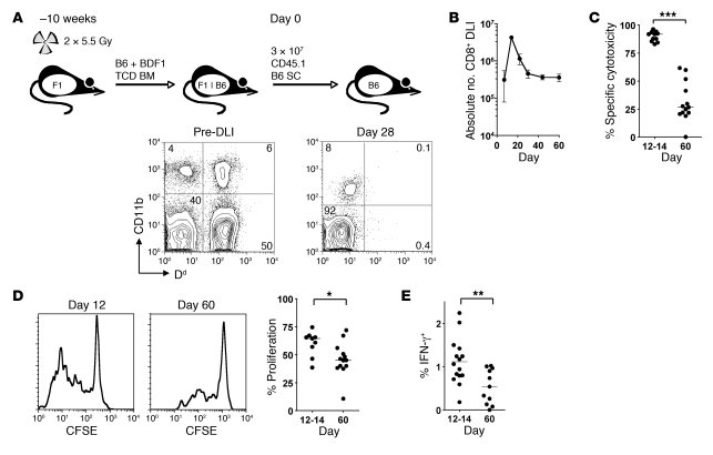
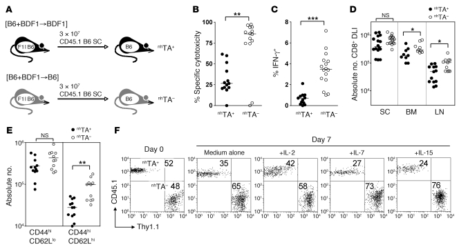
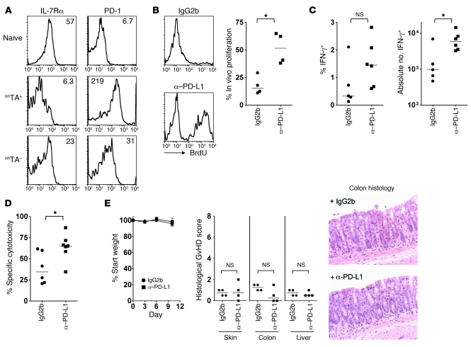
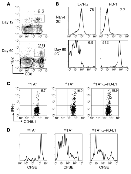
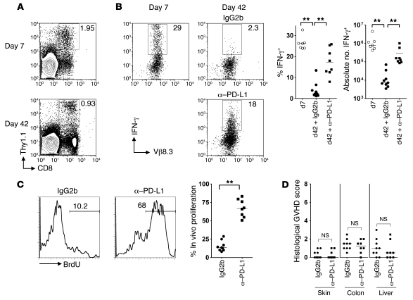
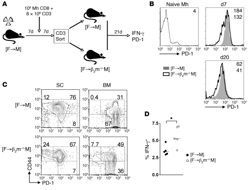
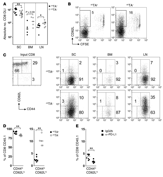
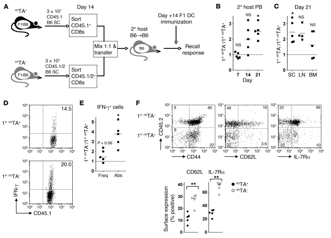
Allogeneic blood or BM transplantation (BMT) is the most commonly applied form of adoptive cellular therapy for cancer. In this context, the ability of donor T cells to respond to recipient antigens is coopted to generate graft-versus-tumor (GVT) responses. The major reason for treatment failure is tumor recurrence, which is linked to the eventual loss of functional, host-specific CTLs. In this study, we have explored the role of recipient antigen expression by nonhematopoietic cells in the failure to sustain effective CTL immunity. Using clinically relevant models, we found that nonhematopoietic antigen severely disrupts the formation of donor CD8+ T cell memory at 2 distinct levels that operate in the early and late phases of the response. First, initial and direct encounters between donor CD8+ T cells and nonhematopoietic cells blocked the programming of memory precursors essential for establishing recall immunity. Second, surviving CD8+ T cells became functionally exhausted with heightened expression of the coinhibitory receptor programmed death-1 (PD-1). These 2 factors acted together to induce even more profound failure in long-term immunosurveillance. Crucially, the functions of exhausted CD8+ T cells could be partially restored by late in vivo blockade of the interaction between PD-1 and its ligand, PD-L1, without induction of graft-versus-host disease, suggestive of a potential clinical strategy to prevent or treat relapse following allogeneic BMT.


Figures

Figure 1
Figure 1. CD8+ T cells transferred to established mixed chimeras show transient cytotoxicity followed by loss of effector function.
(A) Allogeneic chimeras were generated by lethal irradiation of recipient BDF1 (F1) mice followed by reconstitution with T cell–depleted (TCD) B6 and BDF1 BM. 10 weeks later, 3 × 10⁷ CD45.1+ B6 SCs were transferred to established chimeras. A representative example of blood chimerism of mice before and 28 days after DLI is shown using Dd as a marker of BDF1-derived cells. (B) Absolute number of CD45.1+ CD8+ T cells recovered from the spleen at various times following DLI (n = 3 per group per time point). (C) A 1:1 mix of CFSEhi and CFSElo labeled B6 and BDF1 B cells, respectively, were injected intravenously into mice 12 or 60 days after DLI. In vivo cytotoxicity was measured by the disappearance of injected BDF1 B cells relative to B6 B cells after 15 hours. (D) SCs isolated from recipients at 12 or 60 days were CFSE labeled and stimulated for 5 days with irradiated BDF1 SCs. Shown are representative histograms of proliferation of transferred CD8+ T cells and summary data of the percentage of cells that had divided 2 or more times. (E) SCs were restimulated overnight with irradiated BDF1 SCs, and production of IFN-γ by donor CD8+ T cells was measured by intracellular cytokine staining and flow cytometry. Scatter plot indicates the percentage of CD45.1+ CD8+ T cells secreting IFN-γ. *P < 0.05, **P < 0.01, ***P < 0.001, Mann-Whitney test.
Figure 2
Figure 2. Nonhematopoietic antigen disrupts memory function and homeostasis.
(A) nhTA+ [B6+BDF1→BDF1] and nhTA [B6+BDF1→B6] allogeneic chimeras were established and 10 weeks later received 3 × 10⁷ CD45.1+ B6 SCs. (B) In vivo cytotoxicity of CFSE-labeled BDF1 target B cells 60 days after SC transfer. (C) IFN-γ production by CD45.1+ CD8+ T cells recovered from the spleens of recipients 60 days after SC transfer, following overnight stimulation with irradiated BDF1 SCs. (D) Absolute number of DLI-derived CD8+ T cells in the spleen, LN, and BM 60 days after SC transfer. (E) Scatter plots show the absolute number of CD44hiCD62Llo and CD44hiCD62Lhi DLI-derived CD8+ cells recovered from spleen. *P < 0.05, **P < 0.01, ***P < 0.001, Mann-Whitney test. (F) SCs (3 × 10⁷) were transferred from CD45.1+ and Thy1.1+ B6 mice to nhTA+ and nhTA chimeras, respectively. At 60 days after transfer, CD8+ T cells were sorted by magnetic selection, mixed at a 1:1 ratio, and incubated in medium alone or with the indicated cytokines. Plots show starting mixture and the relative survival (as a percentage) of CD8+ T cells from nhTA+ or nhTA hosts after 7 days culture.
Figure 3
Figure 3. Function of exhausted CD8+ T cells can be partially restored by blockade of the PD-1/PD-L1 pathway.
(A) Representative histograms show cell surface expression of IL-7Rα and PD-1 on spleen CD45.1+ CD8+ T cells of nhTA+ or nhTA chimeras 60 days after SC transfer. Numbers indicate MFI: IL-7Rα, nhTA+ 20 ± 2 versus nhTA 61 ± 4, P < 0.001, n = 7 per group; PD-1, nhTA+ 134 ± 16 versus nhTA 29 ± 3, P < 0.001, n = 13–17 per group. (BE) From 48 to 60 days after CD45.1+ B6 SC transfer, mice were administered 0.2 mg anti–PD-L1 or isotype control antibody by i.p. injection every 72 hours and received BrdU in the drinking water during the same period. (B) Representative histograms and summary data showing the percentage of CD45.1+ CD8+ T cells in the spleen that incorporated BrdU. (C) Summary data showing IFN-γ production at 60 days by CD45.1+ CD8+ splenic T cells after overnight stimulation with irradiated BDF1 SCs. (D) In vivo cytotoxicity of CFSE-labeled BDF1 target B cells. (E) Weight change; histological GVHD scores in skin, colon, and liver (scored single-blind); and representative colon histology (original magnification, ×200). Data are representative of 3 independent experiments. *P < 0.05, Mann-Whitney test.
Figure 4
Figure 4. Direct recognition of antigen upon nonhematopoietic antigen is sufficient to drive exhaustion of transferred CD8+ T cells.
B6 SCs (3 × 10⁷) in combination with 2C Tg CD45.1+ CD8+ T cells (2 × 10⁶) were transferred to established nhTA+ chimeras. (A) Contour plots showing frequency of 1B2+ cells as a percentage of CD8+ cells in spleen at 12 and 60 days after transfer to nhTA+ recipients. (B) Histograms show cell surface phenotype of 2C Tg CD8+ T cells (gated using 1B2 clonotypic antibody) from nhTA+ mice at 60 days following transfer. (C) SCs recovered from nhTA+ or nhTA chimeras 60 days after transfer were stimulated overnight with irradiated BDF1 SCs in the presence of isotype control or anti–PD-L1 blocking antibody. Representative dot plots show IFN-γ production by 2C Tg CD8+ T cells (Tg cells were on a CD45.1 background in this experiment). (D) Representative histograms show ex vivo proliferation (in the presence of control or anti–PD-L1 blocking antibody) of 2C CD8+ T cells recovered from the spleen 60 days following transfer to nhTA+ or nhTA chimeras. Data derived from 2 independent experiments with similar results (n = 2–3 mice per group).
Figure 5
Figure 5. Minor H antigen–specific CD8+ T cells recognizing ubiquitously expressed host antigens are susceptible to exhaustion.
B6 female CD3 cells (8 × 10⁶) and Mh Tg Thy1.1+ CD8+ cells (106) were transferred 1 week after lethal irradiation of B6 male mice and reconstitution with female B6 BM. At 36 and 39 days following T cell transfer, mice were administered 0.2 mg anti–PD-L1 blocking antibody or isotype control by i.p. injection and received BrdU in the drinking water during the same period. (A) Representative contour plots showing frequency of Thy1.1+ CD8+ cells as a percentage of SCs. (B) SCs recovered from chimeras at 7 or 42 days after T cell transfer were stimulated overnight with UTY or irrelevant peptide. Representative dot plots and summary data show IFN-γ production by CD8+ Thy1.1+ Mh T cells. Gates set according to irrelevant peptide. (C) Representative histograms and summary data showing the percentage of Thy1.1+ CD8+ T cells incorporating BrdU. (D) Single-blind scoring of histological GVHD in sections taken from skin, colon, and liver of recipient mice. Data derived from 4 independent experiments. **P < 0.01, Mann-Whitney test.
Figure 6
Figure 6. Direct presentation of a minor H antigen by nonhematopoietic cells contributes to exhausted phenotype.
(A) B6 female CD3+ (8 × 10⁶) and Mh Tg female CD8+ (106) cells were transferred 1 week after lethal irradiation of B6 male mice and reconstitution with female B6 BM. 7 days following DLI, T cells were isolated from spleen of primary recipients, and 13 × 106 CD3+ cells (containing 5 × 10⁶ Mh cells) were transferred to [F→M] or [F→β2m–/–M] chimeras on the basis of 1 donor spleen equivalent to 1 secondary recipient (n = 5 per group). Secondary chimeras were set up at the same time as the primary recipients. (B) Representative overlay histograms of surface PD-1 expression upon naive Mh cells and in peripheral blood at 8 and 20 days following secondary transfer. Numbers indicate MFI (top, [F→M] or naive; bottom, [F→β2m–/–M]); day 8, [F→M] 180 ± 7 versus [F→β2m–/–M] 130 ± 7 (P < 0.01); day 20, 58 ± 3 versus 40 ± 1 (P < 0.01). (C) Representative contour plots showing PD-1 and CD44 expression in gated Mh cells within spleen and BM at day 21 following secondary transfer. Figures denote proportion of Mh cells within each gate: spleen, PD-1lo cell frequency [F→M] 14% ± 2% versus [F→ β2m–/–M] 22% ± 1% (P < 0.01); BM, PD-1hiCD44int/lo cell frequency 61% ± 2% versus 43% ± 1% (P < 0.01); BM, PD-1lo cell frequency 1% ± 0.3% versus 7% ± 0.8% (P < 0.01). (D) Summary data of frequency of IFN-γ generation by Mh CD8+ cells following stimulation with UTY peptide. (BD) 1 of 2 independent experiments with similar results. *P < 0.05, Mann-Whitney test.
Figure 7
Figure 7. Nonhematopoietic antigen blocks early generation of postmitotic, CD62L+ CD8+ cells.
(A) CD45.1+ B6 SCs (3 × 10⁷) were transferred to established allogeneic chimeras as in Figure 2. Accumulation of DLI-derived CD8+ T cells in the spleen, LN, and BM in nhTA+ and nhTA recipients 14 days after transfer is shown. (B) Representative dot plots showing CFSE versus CD62L staining in the CD45.1+ CD8+ gated population at day 8. Numbers denote percent CD8+ CFSElo cells that were CD62L+. (C) Representative dot plots showing CD44 and CD62L staining in input CD8+ cells and in CFSElo CD45.1+ CD8+ gated population. (D) Summary data for the same gated population in spleen. Significant differences were also evident in the LN and BM (not shown). (E) nhTA+ chimeras received a transfer of 3 × 10⁷ B6 CD45.1+ SCs followed by i.p. injection of 0.2 mg anti–PD-L1 antibody on days 0, 3, 6, and 9. At 12 days, the proportion of CD45.1+ CD8+ gated cells with a CD44hiCD62Lhi phenotype was determined. Data derived from 3 independent experiments. *P < 0.05, **P < 0.01, Mann-Whitney test.
Figure 8
Figure 8. Nonhematopoietic antigen regulates early memory fate determination.
(A) CD45.1+ or CD45.1/2+ B6 SCs (3 × 10⁷) were transferred to nhTA+ and nhTA chimeras, respectively. 14 days later, CD45.1+ CD8+ T cells from nhTA+ and nhTA chimeras were sorted, mixed at a 1:1 ratio, and transferred to established secondary B6→B6 hosts. After 14 days, secondary hosts were immunized i.p. with 1 × 10⁶ BDF1 LPS-matured DCs, and recall immunity was measured 7 days later. (B) Relative frequency of the 2 transferred CD8+ T cell populations in the blood of secondary hosts at timed intervals following secondary transfer (ratio of CD45.1+CD45.2+ to CD45.1+CD45.2 CD8+ cells). (C) Relative frequency of above populations in the spleen, LN, and BM of secondary hosts at 21 days. (D) Representative plots show IFN-γ production by CD45.1+ CD8+ T cells that were CD45.2 (nhTA+) or CD45.2+ (nhTA). (E) Summary data showing the relative function in terms of the frequency (freq) and absolute number (abs) of CD8+ T cells producing IFN-γ. (B, C, and E) *P < 0.05, Wilcoxon signed rank test relative to expected ratio of 1 (dotted line). (F) Representative dot plots show the cell surface expression of CD44, CD62L, and IL-7Rα on CD8+ CD45.1+ cells 21 days following secondary transfer. Numbers indicate the percentage of gated cells in each quadrant. Scatter plots show the percentage of each transferred cell population expressing cell surface markers. **P < 0.01, Mann-Whitney test.

References

    1. Kolb HJ, Schmid C, Barrett AJ, Schendel DJ. Graft-versus-leukemia reactions in allogeneic chimeras. Blood. 2004;103(3):767–776. doi: 10.1182/blood-2003-02-0342. - DOI - PubMed
    1. Sprangers B, Van Wijmeersch B, Fevery S, Waer M, Billiau AD. Experimental and clinical approaches for optimization of the graft-versus-leukemia effect. Nat Clin Pract Oncol. 2007;4(7):404–414. doi: 10.1038/ncponc0848. - DOI - PubMed
    1. Chakraverty R, et al. An inflammatory checkpoint regulates recruitment of graft-versus-host reactive T cells to peripheral tissues. J Exp Med. 2006;203(8):2021–2031. - PMC - PubMed
    1. Hill GR, Crawford JM, Cooke KR, Brinson YS, Pan L, Ferrara JL. Total body irradiation and acute graft-versus-host disease: the role of gastrointestinal damage and inflammatory cytokines. Blood. 1997;90(8):3204–3213. - PubMed
    1. Xun CQ, Thompson JS, Jennings CD, Brown SA, Widmer MB. Effect of total body irradiation, busulfan-cyclophosphamide, or cyclophosphamide conditioning on inflammatory cytokine release and development of acute and chronic graft-versus-host disease in H-2-incompatible transplanted SCID mice. Blood. 1994;83(8):2360–2367. - PubMed

Publication types