. 2010 Nov;120(11):4111-7.
doi: 10.1172/JCI44425. Epub 2010 Oct 18.

Homologous regions of autoantibody heavy chain complementarity-determining region 3 (H-CDR3) in patients with pemphigus cause pathogenicity

Affiliations

Homologous regions of autoantibody heavy chain complementarity-determining region 3 (H-CDR3) in patients with pemphigus cause pathogenicity

Jun Yamagami et al. J Clin Invest. 2010 Nov.

Abstract

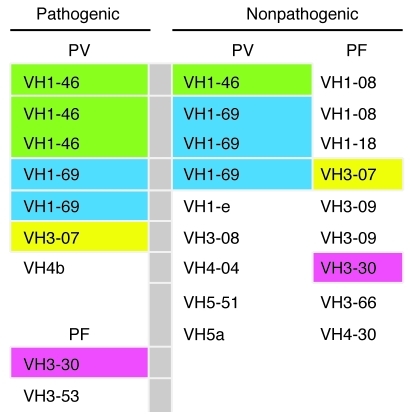
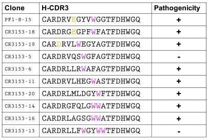
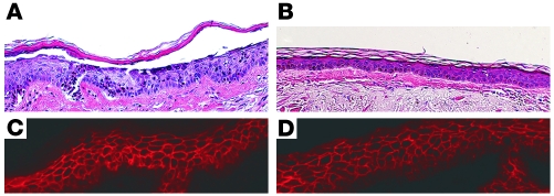
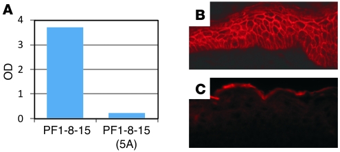
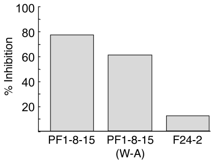
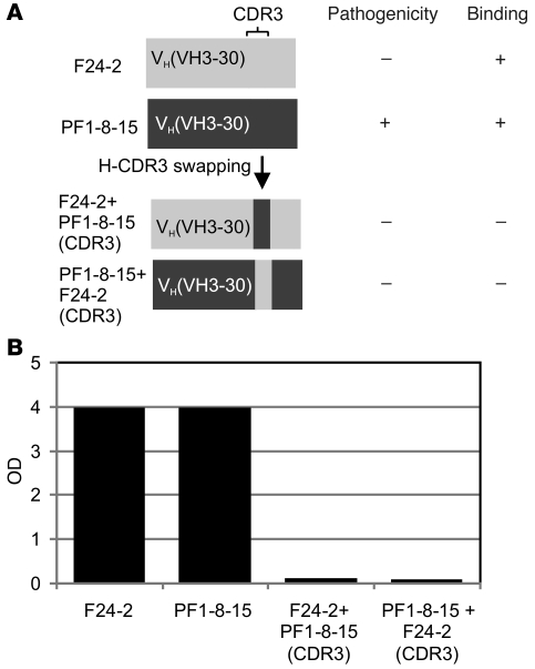
Pemphigus is a life-threatening autoimmune disease in which antibodies specific for desmogleins (Dsgs) cause loss of keratinocyte cell adhesion and blisters. In order to understand how antibodies cause pathogenicity and whether there are commonalities among antibodies in different patients that could ultimately be used to target specific therapy against these antibodies, we characterized Dsg-specific mAbs cloned by phage display from 3 patients with pemphigus vulgaris and 2 with pemphigus foliaceus. Variable heavy chain gene usage was restricted, but similar genes were used for both pathogenic and nonpathogenic mAbs. However, the heavy chain complementarity-determining region 3 (H-CDR3) of most pathogenic, but not nonpathogenic, mAbs shared an amino acid consensus sequence. Randomization of the H-CDR3 and site-directed mutagenesis indicated that changes in this sequence could block pathogenicity but not necessarily binding. In addition, for 2 antibodies with longer H-CDR3s, a tryptophan was critical for pathogenicity but not binding, a result that is consistent with blocking the tryptophan acceptor site that is thought to be necessary for Dsg-mediated adhesion. These studies indicate that H-CDR3 is critical for pathogenicity of a human autoantibody, that a small region (even 1 amino acid) can mediate pathogenicity, and that pathogenicity can be uncoupled from binding in these antibodies.


Figures

Figure 1
Figure 1. VH gene usage of pathogenic and nonpathogenic anti-Dsg mAbs from PV and PF patients.
Colors indicate which genes are shared by pathogenic and nonpathogenic antibodies.
Figure 2
Figure 2. H-CDR3 amino acid sequences from cloned pathogenic mAbs from 3 PV and 2 PF patients.
Bold D, E, and W show D/E-x-x-x-W consensus sequence. YYCAR/S and YYCVR indicate end of VH framework 3. WGQ is beginning of framework 4. AK23 is a mouse monoclonal anti-Dsg3 pathogenic mAb. Common names of some mAbs are in parentheses. Nonbolded orange amino acids are homologous in different antibodies.
Figure 3
Figure 3. H-CDR3 sequences of PF1-8-15 pathogenic anti-Dsg1 mAb and the same mAb with randomized H-CDR3s cloned by binding to Dsg1.
Note that all mAbs have at least one W in the CDR3 (magenta). The original PF1-8-15 and 2 randomized antibodies return the consensus D/E-x-x-x-W sequence.
Figure 4
Figure 4. Normal human skin injection with 2 antibodies derived from PF1-8-15 with randomized H-CDR3s.
CR3153-6 (A and C) causes a superficial epidermal blister typical of PF (A, histology, H&E staining) and binds to the cell surface of epidermal cells (C, immunofluorescence), whereas CR3153-5 does not cause a blister (B) but still binds to the epidermis (D). Injections of human skin with randomized antibodies were done in duplicate with the same results. Original magnification, ×20.
Figure 5
Figure 5. Normal human skin injection of anti-Dsg mAbs.
Histology (H&E) was used to detect pathogenicity (A, C, E, G, I, K, and M) and immunofluorescence to detect mAb binding to the epidermis (B, D, F, H, J, L, and N). The following mAbs were used: PF1-8-15(W–A) (A and B); PF1-8-15(E–D) (C and D); PF1-8-15(E–A) (E and F); PF1-8-15(V–A) (G and H); F24-9(4A) (I and J); F24-9(D–E) (K and L); F24-9(D–A) (M and N). All mAbs bind to the cell surface of epidermal cells and all are pathogenic except PF1-8-15(W–A) and F24-9(4A). Injection of site-directed mutagenesis scFv into normal human skin was repeated 3 times with the same results. Original magnification, ×20.
Figure 6
Figure 6. Characterization of PF1-8-15 anti-Dsg1 antibody and its mutant, PF1-8-15(5A).
ELISA (A) and IIF (C) show the mutant loses its ability to bind Dsg1 but that the parent mAb binds in both ELISA and IIF (B). Original magnification, ×40.
Figure 7
Figure 7. PF 1-8-15(W–A) mutant blocks binding of original PF 1-8-15 to Dsg 1 on ELISA.
Dsg1 ELISA plate was incubated first with the scFv indicated below bars, then with phage expressing PF 1-8-15. ELISA was developed with anti-M13 (phage) antibody. F24-2 is a nonpathogenic anti-Dsg 1 antibody.
Figure 8
Figure 8. Correct context of H-CDR3 in VH is necessary for binding.
Schematic diagram of H-CDR3 swapping between F24-2, a nonpathogenic anti-Dsg1 mAb, and PF1-8-15, a pathogenic mAb (A). ELISA shows that such swapping results in loss of binding to Dsg1 of both mAbs (B).

References

    1. Stanley JR, Amagai M. Pemphigus, bullous impetigo, and staphylococcal scalded skin syndrome. N Engl J Med. 2006;355(17):1800–1810. doi: 10.1056/NEJMra061111. - DOI - PubMed
    1. Mahoney MG, Wang Z, Rothenberger K, Koch PJ, Amagai M, Stanley JR. Explanation for the clinical and microscopic localization of lesions in pemphigus foliaceus and vulgaris. J Clin Invest. 1999;103(4):461–468. doi: 10.1172/JCI5252. - DOI - PMC - PubMed
    1. Stanley JR. Pemphigus and pemphigoid as paradigms of organ-specific, autoantibody-mediated diseases. J Clin Invest. 1989;83(5):1443–1448. doi: 10.1172/JCI114036. - DOI - PMC - PubMed
    1. Schiltz JR, Michel B. Production of epidermal acantholysis in normal human skin in vitro by the IgG fraction from pemphigus serum. J Invest Dermatol. 1976;67(2):254–260. doi: 10.1111/1523-1747.ep12513454. - DOI - PubMed
    1. Hashimoto K, Shafran KM, Webber PS, Lazarus GS, Singer KH. Anti-cell surface pemphigus autoantibody stimulates plasminogen activator activity of human epidermal cells. J Exp Med. 1983;157(1):259–272. doi: 10.1084/jem.157.1.259. - DOI - PMC - PubMed

Publication types