. 2010 Oct 19;1(4):e00183-10.
doi: 10.1128/mBio.00183-10.

Specific control of Pseudomonas aeruginosa surface-associated behaviors by two c-di-GMP diguanylate cyclases

Affiliations

Specific control of Pseudomonas aeruginosa surface-associated behaviors by two c-di-GMP diguanylate cyclases

Judith H Merritt et al. mBio. .

Abstract

The signaling nucleotide cyclic diguanylate (c-di-GMP) regulates the transition between motile and sessile growth in a wide range of bacteria. Understanding how microbes control c-di-GMP metabolism to activate specific pathways is complicated by the apparent multifold redundancy of enzymes that synthesize and degrade this dinucleotide, and several models have been proposed to explain how bacteria coordinate the actions of these many enzymes. Here we report the identification of a diguanylate cyclase (DGC), RoeA, of Pseudomonas aeruginosa that promotes the production of extracellular polysaccharide (EPS) and contributes to biofilm formation, that is, the transition from planktonic to surface-dwelling cells. Our studies reveal that RoeA and the previously described DGC SadC make distinct contributions to biofilm formation, controlling polysaccharide production and flagellar motility, respectively. Measurement of total cellular levels of c-di-GMP in ∆roeA and ∆sadC mutants in two different genetic backgrounds revealed no correlation between levels of c-di-GMP and the observed phenotypic output with regard to swarming motility and EPS production. Our data strongly argue against a model wherein changes in total levels of c-di-GMP can account for the specific surface-related phenotypes of P. aeruginosa.


Figures

FIG 1
FIG 1
Identification of a DGC involved in biofilm formation. (A) Biofilms at 24 h comparing the effects of expressing a His-tagged variant of PA1107 in multicopy plasmid pPA1107 to its vector control (pMQ72) in either the WT or the ΔPA1107 mutant background. The static biofilm assay was performed with M63 G/CAA medium. The bar graph represents quantification of the biofilm assays using OD550 readings of solubilized (CV) solution averaged from four wells of each strain, and the error bars indicate standard deviation. Shown are CV-stained wells (top) and quantification of biofilm formation (bottom). (B) Biofilms at 24 h comparing the WT, the ΔPA1107 and ΔsadC single mutants, and the ΔsadC ΔPA1107 double mutant. Assays were performed as described for panel A.
FIG 2
FIG 2
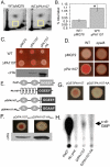
PA1107 is a DGC. (A) Levels of c-di-GMP were quantified after [P]orthophosphate labeling of bacterial cultures, followed by extraction of the nucleotides and analysis via 2D-TLC, for the WT strain expressing a vector control (pMQ78) or a His-tagged variant of PA1107 on a multicopy plasmid (pPA1107). (B) Graphical representation comparing the pixel densities of the c-di-GMP spots from the 2D-TLC plates in panel A normalized to the total pixel density (n = 3). Error bars indicate standard deviations. *, P < 0.05. (C) The WT or ΔPA1107 mutant strain carrying a vector control (pMQ72) or a plasmid expressing a His-tagged variant of WT PA1107 (pPA1107) or a mutant variant altered in the GGEEF domain (pPA1107-AA) was spotted onto CR plates supplemented with arabinose at 0.2%. Plates were imaged after incubation at 37°C for 24 h. This relatively short incubation period is not sufficient for the differences between the WT and ∆roeA mutant strains to become visible. (Bottom) Western blot assay of strains containing the vector control (pMQ72), pPA1107, or pPA1107-AA plasmid probed with anti-His antibody. Loading for the Western blot assay was normalized by OD600. (D) WT or ∆pelA mutant strain carrying a vector control plasmid (pMQ72) or pMQ72 expressing a His-tagged variant of PA1107. CR plates were supplemented with 0.2% arabinose and incubated at 37°C for 24 h. Development of the red color depends on a functional pel locus. (E) Diagram of PleD* and PA1107 domain structure and chimera constructs used for DGC activity assays shown in panels F and H. PleD* (top) contains two N-terminal REC domains (grey ovals labeled “R,” amino acids 3 to 116 and 154 to 265) and a GGDEF domain (box labeled “GGEEF,” amino acids 271 to 452). PA1107 (second from the top) is predicted to have five transmembrane domains (white boxes, between amino acids 34 and 199) followed by a C-terminal GGDEF domain. The PleD*/PA1107 chimeras (bottom two constructs) contain the first 290 amino acids from PleD* and amino acids 202 to 398 from PA1107. The two chimeras contain a small portion of the GGEEF domain from PleD, as well as the complete GGEEF domain of PA1107. Light grey indicates PleD, while the black box indicates the GGEEF domain derived from PA1107. (F) Phenotypes of PleD* chimera constructs in the ΔPA1107 background on CR plates supplemented with 0.2% arabinose. Plates were incubated as described for panel C. (Bottom) Western blot assay of strains containing the pD/Roe603 or pD/Roe-AA603 plasmid. Strains were normalized by OD600 before analysis by Western blotting. (G) Chimeras similar to those described for panel F were built with GST replacing the N terminus of PleD*. Expression of the GST/PA1107 fusion, but not the mutant PA1107-AA variant with a mutation in the GGEEF domain, stimulated CR binding in the WT genetic background. (H) In vitro DGC assay with PleD*/PA1107 chimeras shown in panel F (D/PA1107, D/PA1107-AA), including a PleD* positive control and a no-protein (No prot.) negative control. Reaction products were separated by TLC, followed by exposure to a PhosphorImager plate. The arrow indicates the position of c-di-GMP on the TLC plate.
FIG 3
FIG 3
Mutation of sadC or roeA yields a distinct phenotype. (A) CR plates inoculated with an overnight liquid culture of the WT strain or the ∆roeA, ∆sadC, or ∆pelA mutant strain and incubated for 24 h at 37°C and then at room temperature for 3 days. (B) β-Galactosidase assays of cultures of the indicated strains grown on agar plates with M63 medium supplemented with glucose and CAA for 24 h at 37°C. These are the same conditions used for CR assays, except that the dyes were not added to the growth medium. The relative expression, compared to that of the WT (set at 1), is shown for each strain. Each bar depicts the average of two individual cultures, assayed in triplicate, and the error bars represent the standard deviation (*, P = 0.019 compared to the WT). (C) Swarm plates (0.5% agar) inoculated with liquid cultures of the WT strain or the ∆roeA or ∆sadC mutant strain. Plates were incubated for 16 h at 37°C. (Bottom) Surface area of a plate covered by the swarms (± standard deviation), which was calculated by averaging data from four individual swarm plates. (D) Relative c-di-GMP measurements of the WT (set to a value of 1) and the indicated mutant strains by LC-MS. Assays were performed as described in Materials and Methods. A lowercase letter a above a bar indicates a statistically significant difference from the WT level (P < 0.05). The abbreviation ns indicates that the values below the horizontal bar are not significantly different from each other.
FIG 4
FIG 4
SadC and RoeA are differentially localized. (A) Western blot assays of cellular fractions representing the whole-cell lysate (WCL), cytoplasm (CYT), total membrane (TM), inner membrane (IM), and outer membrane (OM) of the WT strain. SadB, SecY, and OprF are provided as cytoplasmic, IM, and OM controls, respectively, and each protein was detected with a polyclonal antibody. RoeA was detected using anti-His antibody to detect the RoeA-His protein expressed from a plasmid with a PBAD promoter induced with 0.2% arabinose. (B) Control experiments indicating that pSadC-GFP is functional for complementing the ΔsadC mutation in a static biofilm formation assay in the presence of 0.2% arabinose. (C) Control experiments indicating that pRoeA-GFP is functional for complementation of the ΔroeA mutation, performed as described for panel B. (D) Phase-contrast (left) and epifluorescence micrographs (right) of WT P. aeruginosa expressing the vector control pMQ80-GFP (top), pRoeA-GFP (middle), or pSadC-GFP (bottom).
FIG 5
FIG 5
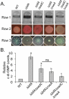
Observed phenotypes do not correlate with total c-di-GMP levels. (A) Biofilm formation (row 1), CR binding (row 2), and swarming (row 3) assays of the strains indicated. CR plates were incubated at 37°C for 24 h, followed by 3 days at room temperature. (B) Relative levels of c-di-GMP in the WT (set to a value of 1) and the mutant strains indicated. Assays were performed as described in Materials and Methods. Letters above bars: a, statistically significant difference from the WT level (P < 0.05); b, statistically significant difference from the ∆bifA mutant (P < 0.05); c, statistically significant difference from the ∆bifA and ∆bifAsadC mutants (P < 0.05). The abbreviation ns indicates that the values below the horizontal bar are not significantly different from each other.

References

    1. Römling U., Gomelsky M., Galperin M. Y. 2005. c-di-GMP: the dawning of a novel bacterial signaling system. Mol. Microbiol. 57:629–639 - PubMed
    1. Miller J. H. 1992. A short course in bacterial genetics. Cold Spring Harbor Laboratory Press, Cold Spring Harbor, NY
    1. Newell P. D., Monds R. D., O’Toole G. A. 2009. LapD is a bis-(3′,5′)-cyclic dimeric GMP-binding protein that regulates surface attachment by Pseudomonas fluorescens Pf0-1. Proc. Natl. Acad. Sci. U. S. A. 106:3461–3466 - PMC - PubMed
    1. Garcia B., Latasa C., Solano C., Garcia-del Portillo F., Gamazo C., Lasa I. 2004. Role of the GGDEF protein family in Salmonella cellulose biosynthesis and biofilm formation. Mol. Microbiol. 54:264–277 - PubMed
    1. Lee V. T., Matewish J. M., Kessler J. L., Hyodo M., Hayakawa Y., Lory S. 2007. A cyclic-di-GMP receptor required for bacterial exopolysaccharide production. Mol. Microbiol. 65:1474–1484 - PMC - PubMed

Publication types

MeSH terms

LinkOut - more resources