

Left ventricular diverticulum mimicking ventricular pseudoaneurysm in an adult
- PMID: 20978576
- PMCID: PMC2953226
Left ventricular diverticulum mimicking ventricular pseudoaneurysm in an adult
Abstract
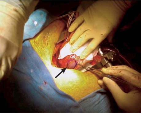
True diverticulum of the left ventricle is very rarely seen in adults: the condition typically occurs in children and can be associated with other anatomic defects that involve the thoracoabdominal midline. Left ventricular diverticulum, which is usually asymptomatic and typically discovered incidentally, can pose a substantial challenge to the surgeon.Herein, we report the case of a 46-year-old man who presented with worsening exertional angina and ST-segment elevation in the inferior electrocardiographic leads. After a stent was deployed in the patient's occluded right coronary artery, left ventriculography revealed outward pouching of the left ventricular inferior wall, suggesting an aneurysm or a contained free-wall rupture. Transesophageal echocardiography showed a sizable defect and a possible intracavitary thrombus. The presumptive diagnosis was a postinfarction subacute pseudoaneurysm of the left ventricle. However, during surgery, we saw no clots, intrapericardial blood accumulation, or perforation. A localized area of thinned muscle in the region of the posterior descending coronary artery was consistent with a ventricular diverticulum. The left ventricular epicardial surface was reinforced with a small bovine pericardial patch. The patient's recovery was uneventful. We discuss the forms of congenital left ventricular diverticulum and offer considerations regarding differential diagnosis.
Keywords: Diagnosis, differential; diverticulum/congenital/diagnosis/physiopathology; heart defects, congenital/diagnosis; heart ventricles/abnormalities; treatment outcome.
Figures


References
-
- Peacock TB. On malformations of the human heart. 2nd ed. London: Churchill & Sons; 1866.
-
- Suilen C, Friedli B, Rutishauser W. Congenital intrathoracic left ventricular diverticulum in an adult. Chest 1990;98(3): 750–1. - PubMed
-
- Walton-Shirley M, Smith SM, Talley JD. Left ventricular diverticulum: case report and review of the literature. Cathet Cardiovasc Diagn 1992;26(1):31–3. - PubMed
-
- Vazquez-Perez J, Gautier M, Mercier JN, Belaisch G, Nouaille J. Diverticulum of the left ventricle (apropos of 3 cases) [in French]. Arch Mal Coeur Vaiss 1969;62(7):922–40. - PubMed
Publication types
MeSH terms
LinkOut - more resources
Full Text Sources
Medical