Review
. 2010 Oct;36(7):723-37.
doi: 10.1055/s-0030-1265289. Epub 2010 Oct 26.

Coagulation abnormalities in the trauma patient: the role of point-of-care thromboelastography

Affiliations
Review

Coagulation abnormalities in the trauma patient: the role of point-of-care thromboelastography

Eduardo Gonzalez et al. Semin Thromb Hemost. 2010 Oct.

Abstract

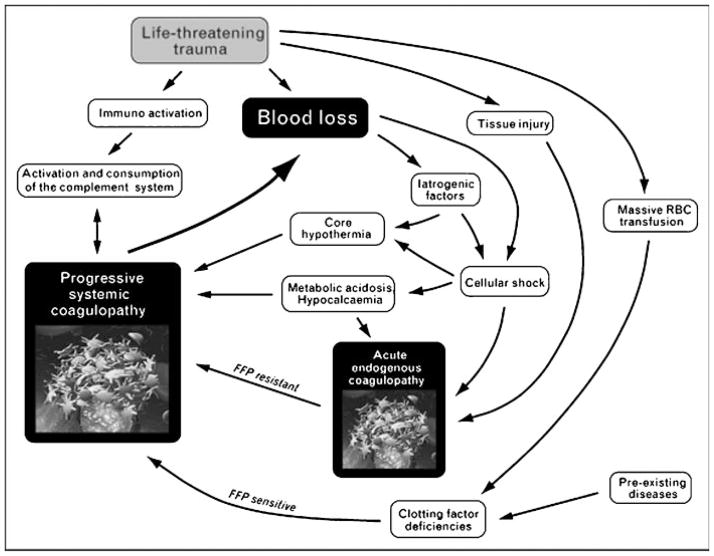
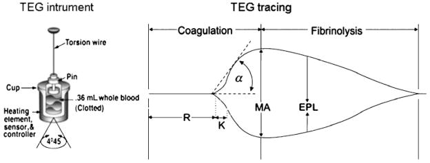
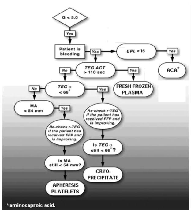
Current recommendations for resuscitation of the critically injured patient are limited by a lack of point-of-care (POC) assessment of coagulation status. Accordingly, the potential exists for indiscriminant blood component administration. Furthermore, although thromboembolic events have been described shortly after injury, the time sequence of post-injury coagulation changes is unknown. Our current understanding of hemostasis has shifted from a classic view, in which coagulation was considered a chain of catalytic enzyme reactions, to the cell-based model (CBM), representing the interplay between the cellular and plasma components of clot formation. Thromboelastography (TEG), a time-sensitive dynamic assay of the viscoelastic properties of blood, closely parallels the CBM, permitting timely, goal-directed restoration of hemostasis via POC monitoring of coagulation status. TEG-based therapy allows for goal-directed blood product administration in trauma, with potential avoidance of the complications resulting from overzealous component administration, as well as the ability to monitor post-injury coagulation status and thromboprophylaxis. This overview addresses coagulation status and thromboprophylaxis management in the trauma patient and the emerging role of POC TEG.


Figures

Figure 1
Figure 1
The updated bloody vicious cycle. This schematic depicts an updated view of the multiple factors contributing to ongoing post-injury coagulopathy. Recent observations suggest that acute endogenous coagulopathy occurs early after traumatic injury. The subsequent development of progressive systemic coagulopathy is time dependent, resulting from persistent hypotension, acidosis, and cellular shock. FFP, fresh-frozen plasma; RBC, red blood cells. Reproduced with permission from Kashuk et al.
Figure 2
Figure 2
Thrombelastography instrument and tracing. The instrument diagram depicts the cuvette where a whole blood sample is placed and the pin attached to a torsion wire. Once the assay is initiated, a tracing is produced and an initial linear segment (zone of precoagulation) extends from the beginning of the test to the formation of the first fibrin strand, causing the tracing to split (split point). The progressive divergence of the tracing reflects the formation of the clot. The R time is reached when the onset of clotting provides enough resistance to produce a 2-mm amplitude reading. The K time is a measurement of the time interval from the R time to the point where fibrin cross-linking provides enough clot resistance to produce a 20-mm amplitude reading. The α angle is the angle formed by the slope of a tangent line traced from the R to the K time measured in degrees. The maximum amplitude, MA, indicates the point at which clot strength reaches its maximum measure in millimeters. Estimated percentage lysis, EPL, is a measure of fibrinolysis and reflects the slow yet progressive decrease in clot strength once MA is reached. Reproduced with permission from Haemonetics Corporation.
Figure 3
Figure 3
Normal and abnormal thrombelastographic patterns. (A) Normal tracing. (B) Prolonged clotting time seen with coagulation factor deficiency or inhibition by anticoagulation. (C) Decreased maximum amplitude (MA), seen during platelet dysfunction or pharmacological inhibition. (D) Increased percentage lysis in a fibrinolytic tracing. (E) Decreased clotting and clotting formation time, elevated α angle, and increased MA represent a hypercoagulable state. Reproduced with permission from Haemonetics Corporation.
Figure 4
Figure 4
Thrombus velocity curve. Sample of a thrombus velocity curve (V curve, in gray), calculated from the first derivative of changes in clot resistance, depicted here over a standard thromboelastographic tracing, showing relationship of delta (R to SP) with thrombus generation parameters. MRTG, maximum rate of thrombus generation; R, reaction time; SP, split point; TMRTG, time to maximum rate of thrombus generation; TTG, total thrombus generation; Δ, delta.
Figure 5
Figure 5
Coagulopathic tracing after injury and subsequent correction via goal-directed therapy. The black thromboelastographic tracing was obtained upon admission and shows delayed clot initiation, decreased rate of clot formation (decreased angle), and poor platelet contribution to clot strength (decreased maximum amplitude). Administration of fresh-frozen plasma, cryoprecipitate, and platelets gradually achieved a normal profile, as seen by the subsequent tracings in gray.
Figure 6
Figure 6
Algorithm of goal-directed approach to post-traumatic coagulopathy. α angle, rate of clot formation; ACA, aminocaproic acid; ACT, activated clotting time; EPL, estimated percentage lysis; FFP, fresh-frozen plasma; G, clot strength; MA, maximum amplitude; r-TEG, rapid thromboelastography; TEG, thromboelastography.
Figure 7
Figure 7
Fibrinolytic tracing. Profound fibrinolysis (tracing in black) seen in hemorrhaging patient after injury. Subsequent correction of coagulation profile is achieved (tracing in gray) after administration of aminocaproic acid.

References

    1. Sauaia A, Moore FA, Moore EE, et al. Epidemiology of trauma deaths: a reassessment. J Trauma. 1995;38:185–193. - PubMed
    1. Kauvar DS, Lefering R, Wade CE. Impact of hemorrhage on trauma outcome: an overview of epidemiology, clinical presentations, and therapeutic considerations. J Trauma. 2006;60(6, Suppl):S3–S11. - PubMed
    1. Cosgriff N, Moore EE, Sauaia A, Kenny-Moynihan M, Burch JM, Galloway B. Predicting life-threatening coagulopathy in the massively transfused trauma patient: hypothermia and acidoses revisited. J Trauma. 1997;42(5):857–861. discussion 861–862. - PubMed
    1. MacLeod JB, Lynn M, McKenney MG, Cohn SM, Murtha M. Early coagulopathy predicts mortality in trauma. J Trauma. 2003;55(1):39–44. - PubMed
    1. Kashuk JL, Moore EE, Millikan JS, Moore JB. Major abdominal vascular trauma—a unified approach. J Trauma. 1982;22(8):672–679. - PubMed

MeSH terms