. 2011 Jan;53(1):230-42.
doi: 10.1002/hep.23999. Epub 2010 Oct 26.

Human T cell microparticles circulate in blood of hepatitis patients and induce fibrolytic activation of hepatic stellate cells

Affiliations

Human T cell microparticles circulate in blood of hepatitis patients and induce fibrolytic activation of hepatic stellate cells

Miroslaw Kornek et al. Hepatology. 2011 Jan.

Abstract

Microparticles (MPs) are small cell membrane vesicles that are released from cells during apoptosis or activation. Although circulating platelet MPs have been studied in some detail, the existence and functional role of T cell MPs remain elusive. We show that blood from patients with active hepatitis C (alanine aminotransferase [ALT] level >100 IU/mL) contains elevated numbers of T cell MPs compared with patients with mild hepatitis C (ALT <40 IU/mL) and healthy controls. T cell MPs fuse with cell membranes of hepatic stellate cells (HSCs), the major effector cells for excess matrix deposition in liver fibrosis and cirrhosis. MP uptake is partly intercellular adhesion molecule 1-dependent and leads to activation of nuclear factor kappa B and extracellular signal-regulated kinases 1 and 2 and subsequent up-regulation of fibrolytic genes in HSCs, down-regulation of procollagen α1(I) messenger RNA, and blunting of profibrogenic activities of transforming growth factor β1. Ex vivo, the induced fibrolytic activity is evident in MPs derived from activated CD4+ T cells and is highest in MPs derived from activated and apoptotic CD8+ T cells. Mass spectrometry, fluorescence-activated cell sorting analysis, and function blocking antibodies revealed CD147/Emmprin as a candidate transmembrane molecule in HSC fibrolytic activation by CD8+ T cell MPs.

Conclusion: Circulating T cell MPs are a novel diagnostic marker for inflammatory liver diseases, and in vivo induction of T cell MPs may be a novel strategy to induce regression of liver fibrosis.


Figures

Figure 1
Figure 1. T cell derived microparticles are found in plasma and elevated in patients with active hepatitis C
(A) Representative FACS analysis of CD3-APC and Annexin V-FITC double positive S100-MP in a plasma sample from a human healthy donor. (B) Relative percentage of circulating CD3 and Annexin V double positive S100-MP from patients with hepatitis C and normal ALT (<40 IU/L; n=4), elevated ALT (>40 IU/L; n=10), or high ALT levels (>100 IU/ml, n=7). (C) CD4/Annexin V and CD8/Annexin double positive, CD14/ Annexin V, CD15/ Annexin V and CD41/Annexin V double positive S100-MP in the plasma of patients with ALT>100 IU/L (n>9) compared to healthy controls and HCV patients with ALT <40 IU/L (n>9). (D) CD8+ S100-MP are ~80 % positive for CD25 (*p<0.05, **p<0.005).
Figure 2
Figure 2. Levels of circulating T cell derived S100-MP correlate with histological grade and stage in patients with hepatitis C
Correlations of plasma CD4+ and CD8+ S100-MP with patients’ biopsies were done as detailed in suppl Methods. Both patients with normal and elevated ALT were included. CD8+ analysis did not work in one patient with Bx stage4.
Figure 3
Figure 3. Characteristics of S100-MP and S10-MP T cell-derived microparticles and demonstration of their fusion with hepatic stellate cell membranes
(A) Ultrastructural analysis of the two subfractions of MP generated from apoptotic Jurkat T cells. Magnification x51,000. (B) Representative forward/sidescatter profiles of events in blood derived S100-MP after addition of beads and intact T cells. (C) FACS analysis demonstrating CD3 receptor transfer from S100-MP to HCS. 2×10⁵ LX-2 cells were incubated with 105 Jurkat T cell-derived S100-MP and CD3 positive LX-2 HSC were quantified after 6 hrs. Unstained HSC and HSC incubated with 0.04μM/mL ST served as controls. (D) Time dependent uptake of CD3 S100-MP by HSC assessed by FACS analysis, demonstrating maximal MP-uptake (15-17%) after 6 hrs; from n=3 events; means±SD; *p=0.003 and **p=0.01. (E) Fluorescence microscopy confirming S100-MP uptake and membrane fusion with HSC. S100-MP were labeled with PKH26 membrane dye and incubated with LX-2 HSC.
Figure 4
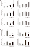
Figure 4. S100-MP from Jurkat T cells elicit antifibrogenic responses in HSC
(A) MMP-1, -3, -9, and -13, TIMP-1 and procollagen α1(I) transcripts were determined by quantitative RT-PCR in LX-2 HSC (2×10⁵ cells each well in 12-well plates) that were incubated with 103 or 5×10⁴ S10-MP or S100-MP from apoptotic Jurkat T cells suspended in 350μL medium for 24hrs. Staurosporine (ST; 0.04μM/mL) or plain medium (medium) served as controls. (B) Induction of fibrolytic and inhibition of fibrogenic genes in TGFβ1 (5ng/mL)-activated HSC when incubated with S100-MP (2×10⁵ MP suspended in 350μL medium) for 24hrs. All experiments were at least performed twice with n=3-4 per group. Results (means±SD) are expressed as arbitrary units relative to beta2-microglobulin mRNA; * p<0.05 vs. medium control.
Figure 5
Figure 5. S100-MP from activated and apoptotic human CD8+ T cells increase MMP and reduce procollagen α1(I) gene expression in HSC in a partly CD54-dependent manner
(A) Transcript levels were determined in LX-2 HSC (2×10⁵ cells/ml per well) incubated with S10-MP or S100-MP (103 or 5×10⁴) from PHA-activated and apoptotic CD8+ T cells for 24hrs by quantitative RT-PCR. ST (0.04μM/mL) or plain medium (medium) served as controls; * p<0.05 vs. medium control. (B) 64% of the S100-MP were CD11a and Annexin V double positive by FACS analysis. (C) HSC were stimulated with TNF-α (10ng/mL) for 0, 4 and 24hrs resulting in a 40% upregulation of CD54 (*p<0.001). (D) Upregulation of CD54 on the surface of HSC by TNF-α facilitated MMP-induction after addition of S100-MP (*p<0.05, **p=0.04, ***p=0.001). (E) HSC were incubated with an CD54 blocking antibody (50μg/mL) or an IgG-matched control antibody for 2 hrs, followed by addition of S100-MP for 24hrs. MMP-3 and -13 transcripts were determined by quantitative PCR. (*p=0.02 and **p=0.046). All experiments were at least performed twice with n=3 per group. Results (means±SD) are expressed as arbitrary units relative to beta2-microglobulin mRNA.
Figure 6
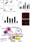
Figure 6. T cell MP engage CD147 (EMMPRIN) on HSC and elicit MMP expression via ERK1/2
(A) FACS analyses of CD147 expression on S100-MP and LX-2 HSC (CD147 positivity 77% and 99%, respectively). (B) CD8+ T cell-derived S100-MP (PHA+ST treatment) were incubated with CD147 blocking antibody (50μg/mL) for 1hr, followed by addition to LX-2 HSC for 24hrs. CD147 blocking significantly decreased MMP-3 and MMP-9 induction as determined by quantitative PCR (by 35%, *p=0.007 and 30%, **p=0.03, respectively). Experiments were performed twice with n=3 per group. Results (means±SD) are expressed as arbitrary units relative to beta2-microglobulin mRNA. (C) Induction of MMP-3 by T cell MP in HSC. Lack of inhibition by the PI3 kinase inhibitor LY294002 (LY, 5μg/ml). Abrogation of MMP-3 induction by the ERK1/2 inhibitor U0126 (U, 5μg/ml), and 50% inhibition by the p38 kinase inhibitor SB203580 (SB, 5μg/ml) and the proteasome (NFkB) inhibitor MG132 (MG, 15μg/ml) (*p=0.02). Comparison to untreated S100-MP stimulated controls. (D) Nuclear translocation of NF-κB p65 in LX-2 HSC exposed to S100-MP from Jurkat T cells for 60min. Representative micrograph from 3 similar experiments. (E) Sketch illustrating the transfer of T cell derived membrane associated molecules including CD147 to HSC membranes via shedded MP. These MP fuse with the HSC membrane which is facilitated by CD54. The transferred receptors can activate novel signaling pathways or auto-/paracrine signaling loops in HSC that favor a switch towards a fibrolytic phenotype via MAP kinase and/or NFkB pathway activation and subsequent induction of MMPs.

References

    1. Schuppan D, Afdhal NH. Liver cirrhosis. Lancet. 2008;371:838–851. - PMC - PubMed
    1. Friedman SL. Mechanisms of hepatic fibrogenesis. Gastroenterology. 2008;134:1655–1669. - PMC - PubMed
    1. Muhanna N, Doron S, Wald O, Horani A, Eid A, Pappo O, Friedman SL, et al. Activation of hepatic stellate cells after phagocytosis of lymphocytes: A novel pathway of fibrogenesis. Hepatology. 2008;48:963–977. - PMC - PubMed
    1. Dienstag JL, Goldin RD, Heathcote EJ, Hann HW, Woessner M, Stephenson SL, Gardner S, et al. Histological outcome during long-term lamivudine therapy. Gastroenterology. 2003;124:105–117. - PubMed
    1. Issa R, Zhou X, Constandinou CM, Fallowfield J, Millward-Sadler H, Gaca MD, Sands E, et al. Spontaneous recovery from micronodular cirrhosis: evidence for incomplete resolution associated with matrix cross-linking. Gastroenterology. 2004;126:1795–1808. - PubMed

MeSH terms