. 2010 Dec;52(6):2148-57.
doi: 10.1002/hep.23915. Epub 2010 Oct 26.

miR-194 is a marker of hepatic epithelial cells and suppresses metastasis of liver cancer cells in mice

Affiliations

miR-194 is a marker of hepatic epithelial cells and suppresses metastasis of liver cancer cells in mice

Zhipeng Meng et al. Hepatology. 2010 Dec.

Abstract

MicroRNAs (miRNAs) are small noncoding RNAs that regulate gene expression by interacting with the 3' untranslated region (3'-UTR) of multiple mRNAs. Recent studies have linked miRNAs to the development of cancer metastasis. In this study, we show that miR-194 is specifically expressed in the human gastrointestinal tract and kidney. Moreover, miR-194 is highly expressed in hepatic epithelial cells, but not in Kupffer cells or hepatic stellate cells, two types of mesenchymal cells in the liver. miR-194 expression was decreased in hepatocytes cultured in vitro, which had undergone a dedifferentiation process. Furthermore, expression of miR-194 was low in liver mesenchymal-like cancer cell lines. The overexpression of miR-194 in liver mesenchymal-like cancer cells reduced the expression of the mesenchymal cell marker N-cadherin and suppressed invasion and migration of the mesenchymal-like cancer cells both in vitro and in vivo. We further demonstrated that miR-194 targeted the 3'-UTRs of several genes that were involved in epithelial-mesenchymal transition and cancer metastasis.

Conclusion: These results support a role of miR-194, which is specifically expressed in liver parenchymal cells, in preventing liver cancer cell metastasis.


Conflict of interest statement

Potential conflict of interest: Nothing to report.

Figures

Fig. 1
Fig. 1
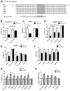
Distribution of miR-194 expression. (A) Dot array of mouse liver miRNAs. (B) Real-time PCR quantification of mature miR-194 in liver tumor and nontumor adjacent tissue control from diethylnitrosamine (100 mg/kg)-induced FXR−/− liver carcinoma models. A Student t test was used to compare the difference between the two groups. *P < 0.05. (C) Real-time PCR quantification of mature miR-194 in human tissue panel.
Fig. 2
Fig. 2
miR-194 was highly expressed in epithelial cells. (A) Quantitative real-time PCR of mature miR-194, miR-122, and miR-21 in isolated mouse hepatic cells: Kupffer cells, stellate cells, and hepatocytes. (B) Levels of miR-194 expression in in vitro cultured mouse primary hepatocytes at different days after perfusion. A Student t test was used to compare the miR-194 expression in each individual day with that of the previous day. *P < 0.05. (C) Levels of miR-194 expression in epithelial or mesenchymal-like liver tumor cell lines. One-way analysis of variance was used to compare miR-194 expression in each individual mesenchymal-like cell line with all the epithelial liver tumor cell lines. *P < 0.05.
Fig. 3
Fig. 3
miR-194 repressed N-cadherin expression in liver mesenchymal-like cancer cells. (A) Comparison of mature miR-194 levels in cells with retroviral expression of miR-194 precursors and controls. A Student t test was used to compare the difference between the two groups. *P < 0.05. CTRL, control. (B) MTS assay of liver cancer cells after retroviral transformation. (C) Western blot analysis of epithelial and mesenchymal cell markers in transformed liver mesenchymal-like cells. The density of the blot was first normalized with a corresponding β-actin loading control and then standardized with protein expression in SK-Hep-1 cells with a control retrovirus infection.
Fig. 4
Fig. 4
miR-194 repressed migration and invasion capacities of liver mesenchymal-like cells. (A) Representative images of morphological changes after overexpression of miR-194 in SK-Hep-1 cells. SK-Hep-1 cells at 0.5 × 10⁶ were plated in a 10-cm culture dish, and images were taken 36 hours after plating. (B) Representative images of invasion assay of SK-Hep-1 cells with miR-194 overexpression. The purple cells were cells that invaded into the lower chamber. (C) Representative images of migration assay of SK-Hep-1 cells with miR-194 overexpression. (D) Quantification of cell number in the invasion assay and the migration assay. A Student t test was used to compare the difference between two groups. *P < 0.05.
Fig. 5
Fig. 5
miR-194 repressed the metastasis of the liver mesenchymal-like cancer cells. Tail vein injection of SK-Hep-1 cells with miR-194 overexpression or controls to SCID mice. (A) Number of tumor foci in SCID mouse livers 4 weeks after injection. A Student t test was used to compare the difference between two groups. *P < 0.05. (B) Representative figures of livers with metastases. (C) Total number of all the metastases in lung determined by hematoxylin-eosin staining and microscopy, and number of the metastases with the width over 0.5 mm. *P < 0.05. (D) Representative hematoxylin-eosin staining of SCID mouse lung. Arrows indicate metastasis foci. CTRL, control.
Fig. 6
Fig. 6
miR-194 targeted several genes involved in EMT or metastasis. (A) Diagram of 3′-UTRs of N-cadherin in different species. (B–F) Luciferase reporter assay of psicheck2.2 vector with 3′-UTR fragments of (B) N-cadherin, (C) RAC1, (D) HBEGF, (E) IGF1R (three seeds with flanking sequences were cloned individually), and (F) other potential miR-194 target genes predicted by TargetScan 5.1. A Student t test was used to compare the difference between two groups. *,#P < 0.05. (G) Luciferase reporter assay using psicheck2.2 vector and miR-194 inhibitors (100 nM) in HepG2 cells. *P < 0.05. (H) Real-time PCR analysis of four miR-194 target genes after transfection of the miR-194 inhibitor. *P < 0.05.

References

    1. Taub R. Liver regeneration: from myth to mechanism. Nat Rev Mol Cell Biol. 2004;5:836–847. - PubMed
    1. Tung-Ping Poon R, Fan ST, Wong J. Risk factors, prevention, and management of postoperative recurrence after resection of hepatocellular carcinoma. Ann Surg. 2000;232:10–24. - PMC - PubMed
    1. Huber MA, Kraut N, Beug H. Molecular requirements for epithelial-mesenchymal transition during tumor progression. Curr Opin Cell Biol. 2005;17:548–558. - PubMed
    1. Bartel DP. MicroRNAs: genomics, biogenesis, mechanism, and function. Cell. 2004;116:281–297. - PubMed
    1. Gregory PA, Bert AG, Paterson EL, Barry SC, Tsykin A, Farshid G, et al. The miR-200 family and miR-205 regulate epithelial to mesenchymal transition by targeting ZEB1 and SIP1. Nat Cell Biol. 2008;10:593–601. - PubMed

Publication types