. 2010 Oct;28(10):1829-38.
doi: 10.1002/stem.512.

Allogeneic periodontal ligament stem cell therapy for periodontitis in swine

Affiliations

Allogeneic periodontal ligament stem cell therapy for periodontitis in swine

Gang Ding et al. Stem Cells. 2010 Oct.

Abstract

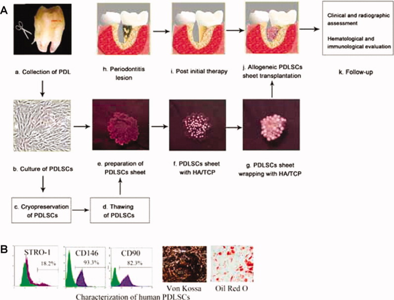
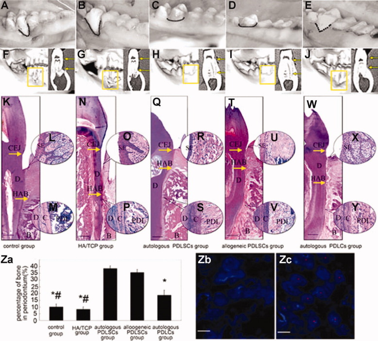
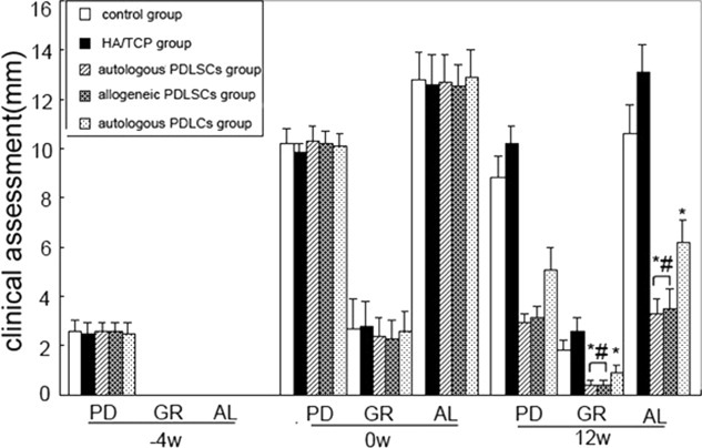
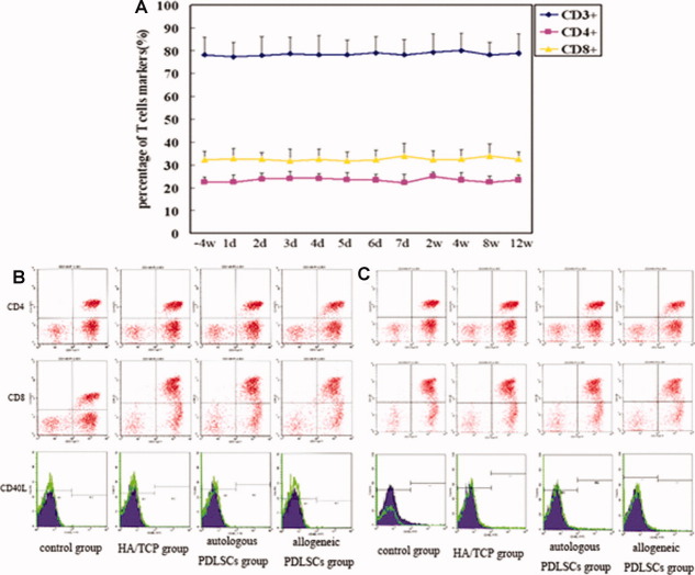
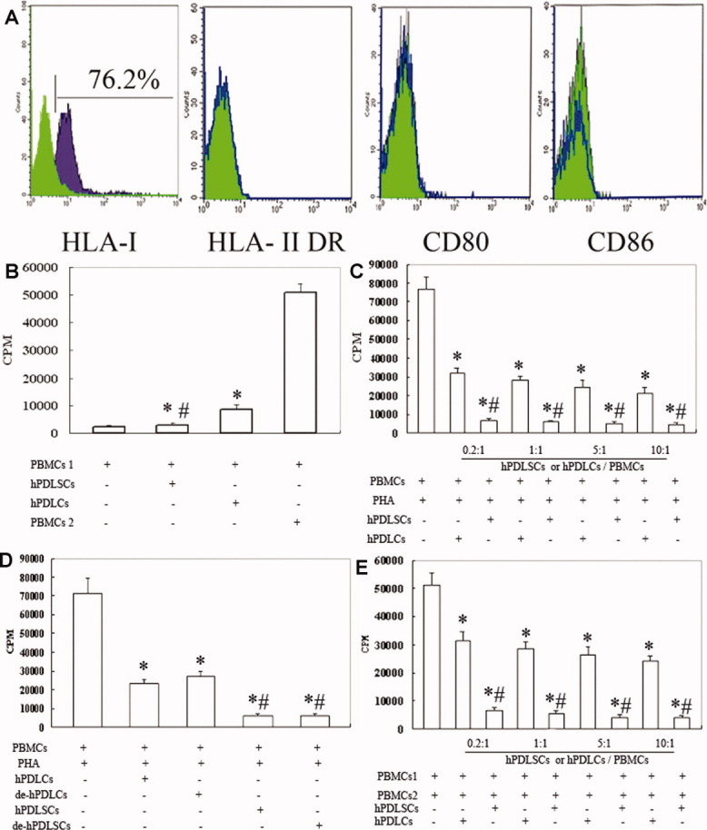
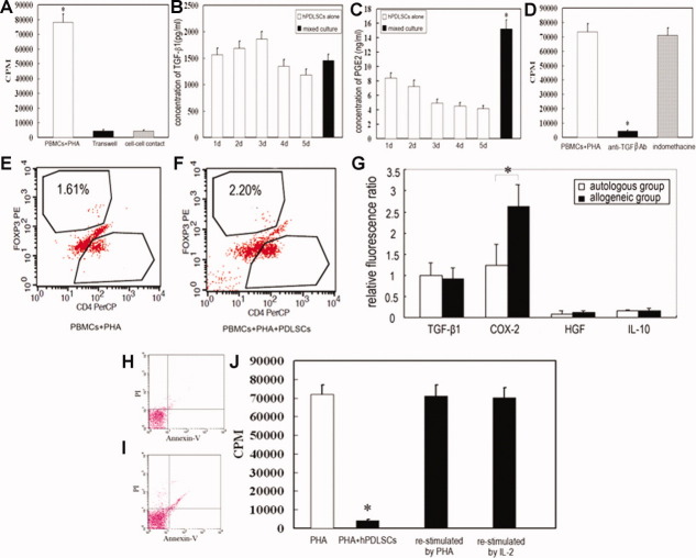
Periodontitis is one of the most widespread infectious diseases in humans. It is the main cause of tooth loss and associated with a number of systemic diseases. Until now, there is no appropriate method for functional periodontal tissue regeneration. Here, we establish a novel approach of using allogeneic periodontal ligament stem cells (PDLSCs) sheet to curing periodontitis in a miniature pig periodontitis model. Significant periodontal tissue regeneration was achieved in both the autologous and the allogeneic PDLSCs transplantation group at 12 weeks post-PDLSCs transplantation. Based on clinical assessments, computed tomography (CT) scanning, and histological examination, there was no marked difference between the autologous and allogeneic PDLSCs transplantation groups. In addition, lack of immunological rejections in the animals that received the allogeneic PDLSCs transplantation was observed. Interestingly, we found that human PDLSCs fail to express human leukocyte antigen (HLA)-II DR and costimulatory molecules. PDLSCs were not able to elicit T-cell proliferation and inhibit T-cell proliferation when stimulated with mismatched major histocompatibility complex molecules. Furthermore, we found that prostaglandin E2 (PGE2) plays a crucial role in PDLSCs-mediated immunomodulation and periodontal tissue regeneration in vitro and in vivo. Our study demonstrated that PDLSCs possess low immunogenicity and marked immunosuppression via PGE2-induced T-cell anergy. We developed a standard technological procedure of using allogeneic PDLSCs to cure periodontitis in swine.


Figures

Figure 1
Figure 1
A standard procedure for the application of allogeneic PDLSCs in the treatment of periodontitis and characterization of PDLSCs. (A): (a) Collection of PDL. Normal impacted third molars were collected from patients aged 18–28 years. PDL was gently separated from the root surface. (b) Culture of PDLSCs. PDLSCs were isolated as previously reported. Fifteen days after primary culture, the number of PDLSCs from one impacted third molar was approximately 4.90 ± 0.34 × 10⁵ (P0; n = 10). Following another 15-day culture, the number of PDLSCs (P3) increased to 8.86 ± 0.46 × 10⁶ (n = 10). (c) Cryopreservation of PDLSCs. Third-passage PDLSCs were cryopreserved with 10% DMSO and 90% FBS and stored in liquid nitrogen. (d) PDLSCs thawing. After thawing, PDLSCs were examined for mycoplasma, bacteria, colony-forming efficiency, profiles of mesenchymal stem cell markers, and karyotype. (e) Preparation of PDLSC sheet. Three different cell amounts (1 × 10⁵, 1 × 10⁶, and 2 × 10⁶, n = 3) were cultured for 12–15 days in 100-mm culture dishes; cell sheets formed in the 1 × 10⁶ and 2 × 10⁶ groups but not in the 1 × 10⁵ group. Therefore, 1 × 10⁶ PDLSCs were seeded into 100-mm culture dishes for 15 days. (f) Forty milligrams of HA/TCP was added to the cultures. (g) The whole view of the PDLSC sheet with HA/TCP. (h) Buccal view of periodontitis lesion. After initial periodontal therapy (i), two allogeneic PDLSC sheets with HA/TCP were transplanted to a periodontal lesion sized 3 mm × 5 mm × 7 mm (j). (k) Follow-up included clinical and radiographic assessments and hematological and immunological evaluations. (B): PDLSCs were positive for STRO-1, CD146, CD90 and could advance into osteogenic and adipogenic differentiation under inductive medium. Abbreviations: HA/TCP, hydroxyapatite/tricalcium phosphate; PDL, periodontal ligament; PDLSC, periodontal ligament stem cell.
Figure 2
Figure 2
Regeneration of periodontitis defects mediated by allogeneic miniature pig PDLSCs (pPDLSCs). (A–E): Intraoral photographs indicated that 12 weeks after transplantation, autologous (C) or allogeneic (D) pPDLSC-mediated periodontal tissue regeneration was close to normal level. Only limited periodontal tissues were regenerated in the control group (A), HA/TCP group (B), and partially regenerated periodontal tissues in pPDLCs group (E). CT images showed that limited bone formation was found in the control group (F) and the HA/TCP group (G) 12 weeks after pPDLSC transplantation. Autologous pPDLSCs (H) or allogeneic pPDLSCs (I) mediated nearly complete alveolar bone regeneration. (K–Y): HE staining showed new periodontal tissue regeneration in the periodontal defect area in autologous (Q) and allogeneic (T) pPDLSC groups. However, deep periodontal pockets and shortages of new bone and periodontal ligament fibers remained in the control (K) and HA/TCP groups (N). The alveolar bone heights in the autologous (Q) and allogeneic (T) pPDLSC groups were much larger than those in the control group (K), the HA/TCP group (N), and PDLCs group (W). Much thicker sulcular epithelia and enlarged epithelial pegs were evident in the control group (L), the HA/TCP group (O), and PDLCs group (X) compared with the autologous (R) and allogeneic (U) pPDLSC groups. Sharpy's fibers formed in the autologous (S) and allogeneic (V) pPDLSC groups, but typical Sharpy's fibers were not found in the control group (M), the HA/TCP group (P), or PDLCs group (Y). (Z): (a) The percentage of periodontal bone in the autologous and allogeneic pPDLSC groups was significantly higher than those of the control group, HA/TCP group, and PDLCs group. (b) No positive staining of Y chromosomes was detected in the autologous pPDLSC group. (c) Positive staining of Y chromosomes was found in alveolar bone close to the PDL in the allogeneic pPDLSC group, indicating that regenerated periodontal tissues differentiated from allogeneic pPDLSCs. “D” indicates dentin, “C” indicates cementum, and “B” indicates bone (K–W). (A, F, K, L, M): Control group without HA/TCP implantation, (B, G, N, O, P): HA/TCP group, (C, H, Q, R, S): autologous pPDLSC group, (D, I, T, U, V): allogeneic pPDLSC group, and (E, J, W, X, Y): autologous PDLCs group. The figures in the upper-right section of (F–J) are the coronal images of miniature pigs. Scale bar = 1 mm (K, N, Q, T, W); Scale bar = 5 μm ([Z], b, c). #, p < .01, compared with autologous PDLCs groups; *, p < .01, compared with autologous and allogeneic PDLSCs groups. Abbreviations: CEJ, cemeto-enamel junction; HA/TCP, hydroxyapatite/tricalcium phosphate; HAB, height of alveolar bone; PDL, periodontal ligament; PDLSC, periodontal ligament stem cell; SE, sulcular epithelium.
Figure 3
Figure 3
Clinical assessment of regeneration of periodontal tissues. At −4 week and 0 week, there was no significant difference among the five groups. However, 12 weeks post-transplantation, the PD, GR, and AL were significantly improved in autologous pig PDLSCs (pPDLSCs) or allogeneic pPDLSCs group than those in the control group, HA/TCP group, and autologous PDLCs group. *, p < .01, compared with autologous and allogeneic PDLSCs groups; #, p < .01, compared with autologous PDLCs groups. Abbreviations: AL, attachment loss; GR, gingival recession; HA/TCP, hydroxyapatite/tricalcium phosphate; PD, probing depth; PDLSC, periodontal ligament stem cell.
Figure 4
Figure 4
Dynamic evaluation of immunological parameters. (A): There were no significant differences in the indicated time points regarding the percentage of CD3+, CD4+, and CD8+ T cells in the allogeneic pig PDLSC transplantation group. (B): The expression of CD4+ and CD8+ T cells, as well as CD40L, a marker of activated T cells, were nearly identical among the four test groups 3 days post-transplantation. (C): The percentage of CD4+ and CD8+ T cells and the expression of CD40L were also nearly identical among the four groups 12 weeks after transplantation. Abbreviations: HA/TCP, hydroxyapatite/tricalcium phosphate; PDLSC, periodontal ligament stem cell.
Figure 5
Figure 5
PDLSCs inhibit T-cell proliferation. (A): hPDLSCs were stained for immunological markers and analyzed by flow cytometry, revealing positive expression of HLA-I but negative expression of HLA-II DR, CD80, and CD86. Data represent five independent experiments. (B): Coculture of T cells with hPDLSCs showed that hPDLSCs failed to stimulate allogeneic T-cell proliferation. *, p < .01, compared with group of coculture of two allogeneic PBMCs; #, p < .01, compared with PDLCs groups. (C): T-cell proliferation stimulated by PHA in the presence or absence of hPDLSCs; the resulting inhibition was dose-dependent. *, p < .01, compared with PHA-stimulated PBMCs group; #, p < .01, compared with PDLCs groups. (D): T-cell proliferation induced by PHA was inhibited by delayed addition of hPDLSCs. *, p < .01, compared with PHA-stimulated PBMCs group; #, p < .01, compared with PDLCs groups. (E): A two-way mixed lymphocyte reaction (MLR) was inhibited by hPDLSCs in a dose-dependent manner.*: p < .01, compared with MLR group; #, p < .01, compared with PDLCs groups. Data are presented as mean ± SD of triplicates of six independent experiments. Abbreviations: CPM, counts per minute; HLA, human leukocyte antigen; hPDLC, heterogenic periodontal ligament cell; hPDLSC, human periodontal ligament stem cell; PBMC, peripheral blood mononuclear cell; PHA, phytohemagglutinin.
Figure 6
Figure 6
Prostaglandin E2 (PGE2) is the key inhibitor of T-cell proliferation mediated by PDLSCs. (A): T-cell proliferation was significantly inhibited when cocultured with human PDLSCs (hPDLSCs) in the Transwell culture system and in cell-cell contact culture. (B): TGF-β1 levels were similar between hPDLSCs alone and the coculture of hPDLSCs and PHA-induced T-cells. (C): The concentration of PGE2 significantly increased in the coculture of hPDLSCs and PHA-induced T cells. (D): The neutralizing monoclonal antibody against TGF-β failed to restore T-cell proliferation, while the PGE2 inhibitor indomethacine restored T-cell proliferation. (E, F): The percentage of Foxp3+ T cells in coculture of PBMCs and PHA (E) was similar to that in coculture of PBMCs, PHA, and hPDLSCs (F). (G): The expression of COX2 was significantly higher in regenerated periodontal tissues of the allogeneic PDLSCs group than in the autologous PDLSCs group 2 weeks after transplantation, whereas TGF-β1, HGF, and IL-10 levels did not change (n = 4). (H, I): T-cell apoptosis rates were similar between PHA-stimulated T cells (I) and a coculture group with hPDLSCs (H). (J): Inhibition of T cell proliferation mediated by hPDLSCs was restored when T cells were restimulated by PHA or IL-2. Data are presented as mean ± SD of triplicates of six independent experiments. *, p < .01. Abbreviations: CPM, counts per minute; HGF, hepatocyte growth factor; hPDLSC, human periodontal ligament stem cell; PBMC, peripheral blood mononuclear cell; PDLSC, periodontal ligament stem cell; PHA, phytohemagglutinin; TGF-β1, transforming growth factor β1.

References

    1. Kinane DF, Marshall GJ. Periodontal manifestations of systemic disease. Aust Dent J. 2001;46:2–12. - PubMed
    1. Kitamura M, Nakashima K, Kowashi Y, et al. Periodontal tissue regeneration using fibroblast growth factor-2: Randomized controlled phase II clinical trial. Plos One. 2008;3:e2611–e2622. - PMC - PubMed
    1. Nygaard-Østby P, Bakke V, Nesdal O, et al. Periodontal healing following reconstructive surgery: Effect of guided tissue regeneration using a bioresorbable barrier device when combined with autogenous bone grafting. A randomized-controlled Trial 10-year follow-up. J Clin Periodontol. 2010;37:366–373. - PubMed
    1. Lee JS, Wikesjö UM, Jung UW, et al. Periodontal wound healing/regeneration following implantation of recombinant human growth/differentiation factor-5 in a beta-tricalcium phosphate carrier into one-wall intrabony defects in dogs. J Clin Periodontol. 2010;37:382–389. - PubMed
    1. Villar CC, Cochran DL. Regeneration of periodontal tissues: Guided tissue regeneration. Dent Clin North Am. 2010;54:73–92. - PubMed

Publication types