Review
. 2010 Oct;217(4):276-88.
doi: 10.1111/j.1469-7580.2010.01281.x.

Renewed focus on the developing human neocortex

Affiliations
Review

Renewed focus on the developing human neocortex

Gavin Clowry et al. J Anat. 2010 Oct.

Abstract

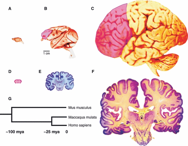
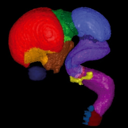
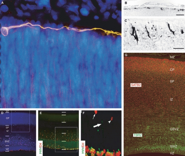
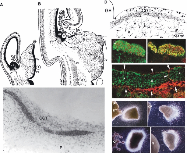
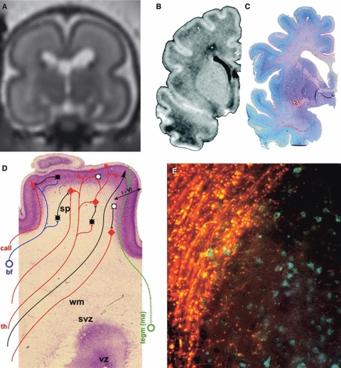
Many specifically human psychiatric and neurological conditions have developmental origins. Rodent models are extremely valuable for the investigation of brain development, but cannot provide insight into aspects that are specifically human. The human brain, and particularly the cerebral cortex, has some unique genetic, molecular, cellular and anatomical features, and these need to be further explored. Cortical expansion in human is not just quantitative; there are some novel types of neurons and cytoarchitectonic areas identified by their gene expression, connectivity and functions that do not exist in rodents. Recent research into human brain development has revealed more elaborated neurogenetic compartments, radial and tangential migration, transient cell layers in the subplate, and a greater diversity of early-generated neurons, including predecessor neurons. Recently there has been a renaissance of the study of human brain development because of these unique differences, made possible by the availability of new techniques. This review gives a flavour of the recent studies stemming from this renewed focus on the developing human brain.


Figures

Fig. 1
Fig. 1
Cerebral hemispheres of the mouse (A), macaque monkey (B) and human (C) drawn at approximately the same scale to convey the overall difference in the size and elaboration of the cerebral cortex. The pink overlay indicates the area of the prefrontal cortex that has no counterpart in mouse. The coronal sections of the cerebral hemispheres of the same species (D–F) illustrate the relatively small increase in the thickness of the neocortex compared with a large difference in surface of approximately 1 : 100 : 1000 in mouse, macaque monkey and human, respectively. (G) The time-scale of phylogenetic divergence of Mus musculus, Maccaca mulata and Homo sapiens based on the DNA sequencing data. Modified from Rakic (2009). Original panels are from the Comparative Mammalian Brain Collection: http://www.mirrorservice.org/sites/brainmuseum.org/index.html.
Fig. 2
Fig. 2
Subdivisions of the developing human brain in a 3D virtual model of an embryo at Carnegie Stage 22 (approximately 7–8 gestational weeks). Different regions have been defined and presented in different colours: pallium, red; subpallium, orange; hypothalamus, brown; diencephalon, shades of green; mesencephalon, blue; metencephalon, shades of purple; myelencephalon, magenta; spinal cord, dark red; dorsal root ganglia, blue. For reference the eye has been painted dark blue, and the inner ear yellow. Figure provided by Dr Janet Kerwin, Newcastle University (http://www.HUDSEN.org).
Fig. 3
Fig. 3
Cerebral cortical germinal zone during neurogenesis and the first postmitotic neurons of the human brain. (A) The first postmitotic neurons, the predecessor neurons in the primordial plexiform layer (PPL) of the human cerebral cortex, were revealed with TU-20 immunostaining (orange) in a coronal section of a Carnegie Stage 13 (4–4.5 gestational weeks) dorsal cortex. The predecessor cells and their tangentially oriented processes populate the PPL in the telencephalon, with a clear basal-to-dorsal density gradient prior to local cortical neurogenesis. They are not immunoreactive to reelin. Reelin-expressing Cajal-Retzius cells in mouse embryo cortex at embryonic day 14 (B) and in human fetus cortex at 21 gestational weeks (C). Cajal-Retzius cells increase in numbers and morphological complexity in mammals. Scale bars: 40 μm (B), 20 μm (C). (D–F) At 11 gestational weeks, the human cortical germinal zone consists of the ventricular and subventricular zones (VZ and SVZ, respectively). The intermediate zone (IZ), subplate (SP), cortical plate (CP) and marginal zone (MZ) were also present in the developing cortical wall as revealed by bisbenzimide (blue). H3+ cells were prominent in the VZ and extraventricular compartments. Expression of H3 and 4A4 was studied with double immunohistochemistry (E) and co-expression in these cells was confirmed by confocal microscopy (F), H3 (red) and 4A4 (green). 4A4 immunoreactivity was primarily restricted to the VZ/SVZ although double-positive cells were present in the apical portion of the IZ. The arrowhead indicates a single-labelled pH3-immunorective profile in the SVZ; the small arrow depicts a similar profile in the IZ. A pial-directed process originating from a radial glia in the VZ is indicated by a thick arrow. (G) Further compartmentalization of the germinal zone to internal SVZ (ISVZ) and outer SVZ (OSVZ) is apparent at 12 gestational weeks on a TBR2- and SATB2-immunostained cerebral cortical slice. TBR2, expressed by intermediate neuronal progenitor cells, marks out the proliferative zones, whereas SATB2, expressed by some postmitotic neurons, marks out the IZ, SP, CP and MZ. All parts of this figure are reproduced with permission: A is from Bystron et al. (2006); B,C from Molnár et al. (2006); D–F from Carney et al. (2007); and G was kindly provided by Bui Kar Ip (Newcastle University).
Fig. 4
Fig. 4
A transient, human-specific structure, the corpus gangliothalamicus (CGT), is situated close to the telo-diencephalic junction, between the telecephalic ganglionic eminence (GE) and dorsal thalamus (DT) in human fetal cerebrum. Semi-diagrammatic drawings of the human thalamus at 10 (A) and 24 (B) gestational weeks showing early genesis of thalamic neurons from the local proliferative ependyma, now called ventricular zone and their migration from the GE via the corpus gargliothalamises (CGT) to the pulvinar (P) at early and late fetal stages, respectively. (C) CGT at the surface of the pulvinar (P) in a human 18 gestational week fetus stained with Cresyl violet. (D) Drawing of the Golgi-impregnated images of migrating neurons in the CGT and transitional forms to neurons beneath. (E) Neurons in the CGT double-immunostained with GE-specific marker Dlx 1/2 and Tuj1 and GABA are indicated with arrows and asterisks, respectively. (F) Use of organotypic cultures to assay the attractive effect of the human DT and the repellent effect of the mouse and human choroid plexus (CP) and mouse subthalamic nucleus (S) on neurons migrating from the explants of the human GE. A–D from Rakic & Sidman (1969); E,F from Letinic & Rakic (2001). 3v, third ventricle; C, caudate nucleus; Cl, clausfrum; CM, centrum medianum; GE, ganglionic eminence; GP, globus pallidus; LV, lateral ventricle; H, hippocampus.
Fig. 5
Fig. 5
Magnetic resonance imaging (MRI) and histological investigations on subplate neurons in human and development of early human cortical circuits. (A) In-utero MRI of the fetal subplate. The layers of the hemisphere are clearly seen on the T2-weighted single-shot images in the coronal plane acquired in a fetus at 23 gestational weeks using a 1.5 T scanner. The subplate is seen as high signal intensity, reflecting its hydrophilic extracellular matrix. At this stage of development the subplate is thicker than the cortex. The developing white matter has a low signal intensity band reflecting increased cellular content (reproduced with permission from Wang et al. 2010b this issue). Low-power view of T1-weighted MRI (B) and Periodic acid-Schiff (PAS)-stained coronal sections (C) through the brains of premature newborns at 36 gestational weeks demonstrating the gradual dissolution of the subplate zone. Reproduced with permission from Kostović et al. (2002). (D) Schematic summary of the neuronal elements involved in early human cortical circuits superimposed on a Nissl-stained section of a 34 gestational week preterm human infant. This period is characterized by the co-existence of transient circuitry in the subplate zone and elaboration of permanent (sensory-driven) circuitry in the cortical plate. Note the initial six layers (I–VI) and the increase in GABAergic neurons (white circles) in the cortical plate. GABAergic neurons, black circles; glutamatergic neurons, red diamond; cortical plate neurons, violet. Afferents from the basal forebrain (bf, blue), monoaminergic brain stem nuclei (tegm, green) and thalamus (th, red). call, callosal fibers; wm, white matter; svz, subventricular zone; vz, ventricular zone. Reproduced with permission from Kostović & Judaš (2007). (E) Neuronal heterotopias are seen in various pathologies and are associated with intractable epilepsy. Studies using histological and carbocyanine dye (1,1′-dioctadecyl-3,3,3′,3′-tetramethylindocarbocyanine perchlorate) tracing techniques in selected cases of subcortical or periventricular nodular heterotopia revealed abnormally differentiated neurons with altered connectivity. Bisbenzimide labelling (blue) reveals the cells and boundaries of the nodules and DiI labelling (orange) demonstrates the vast majority of labelled fibres coursing around but not within the nodules. Some fibres are extending across the margin of the nodule and have punctate termini close to cell bodies within the nodules. Reproduced with permission from Hannan et al. (1999).
Fig. 6
Fig. 6
Age-associated interindividual variations in cortical development have been studied with non-invasive magnetic resonance imaging. Postprocessing of high-quality T1- and T2-weighted images enabled the segmentation of the cortex in large regions across both hemispheres of the brain. Dedicated postprocessing tools enabled the quantitative study of the variations with increases in the cortex (a) and decreases in white matter (b) for the left (L, up) and right (R, down) hemispheres in the preterm newborn. The first two rows show statistical T-maps (colour coded blue to red) that are superposed to the 3D averaged cortical surface. Significant clusters showing ‘apparent increases’ in cortex (a) and ‘apparent decreases’ in white matter (b). (c) The clusters for the cortex (blue) and white matter (red) had considerable overlay (black). Note that the right hemisphere shows larger regions of age-associated variations in comparison with the left. In the third row, statistical T-maps are presented with the most significant clusters along the horizontal, coronal and parasagittal planes. In the lower two rows interhemispherical asymmetries are shown in the preterm group; statistical T-maps are superposed to the 3D averaged cortical surface in the significant clusters showing asymmetries in cortex (a) and white matter (b), for the left (L, up) and right (R, down) hemispheres. The clusters (c) for cortex (blue) and white matter (red) are mainly overlying (black). Reproduced with permission from Dubois et al. (2010).

References

    1. Abrahams BS, Tentler D, Perederiy JV, et al. Genome wide analyses of human perisylvian cerebral cortical patterning. Proc Natl Acad Sci USA. 2007;104:17849–17854. - PMC - PubMed
    1. Ang ES, Jr, Gluncic V, Duque A, et al. Prenatal exposure to ultrasound waves impacts neuronal migration in mice. Proc Natl Acad Sci USA. 2006;103:12903–12910. - PMC - PubMed
    1. Ayoub AE, Kostovic I. New horizons for the subplate zone and its pioneering neurons. Cereb Cortex. 2009;19:1705–1707. - PubMed
    1. Basu A, Graziadio S, Smith M, et al. Developmental plasticity connects visual cortex to motoneurons after stroke. Ann Neurol. 2010;67:132–136. - PubMed
    1. Batista-Brito R, Machold R, Klein C, et al. Gene expression in cortical interneuron precursors is prescient of their mature function. Cereb Cortex. 2008;18:2306–2317. - PMC - PubMed

LinkOut - more resources