Review
. 2010 Oct;217(4):418-28.
doi: 10.1111/j.1469-7580.2010.01280.x.

Neuroimaging of cortical development and brain connectivity in human newborns and animal models

Affiliations
Review

Neuroimaging of cortical development and brain connectivity in human newborns and animal models

Gregory A Lodygensky et al. J Anat. 2010 Oct.

Abstract

Significant human brain growth occurs during the third trimester, with a doubling of whole brain volume and a fourfold increase of cortical gray matter volume. This is also the time period during which cortical folding and gyrification take place. Conditions such as intrauterine growth restriction, prematurity and cerebral white matter injury have been shown to affect brain growth including specific structures such as the hippocampus, with subsequent potentially permanent functional consequences. The use of 3D magnetic resonance imaging (MRI) and dedicated postprocessing tools to measure brain tissue volumes (cerebral cortical gray matter, white matter), surface and sulcation index can elucidate phenotypes associated with early behavior development. The use of diffusion tensor imaging can further help in assessing microstructural changes within the cerebral white matter and the establishment of brain connectivity. Finally, the use of functional MRI and resting-state functional MRI connectivity allows exploration of the impact of adverse conditions on functional brain connectivity in vivo. Results from studies using these methods have for the first time illustrated the structural impact of antenatal conditions and neonatal intensive care on the functional brain deficits observed after premature birth. In order to study the pathophysiology of these adverse conditions, MRI has also been used in conjunction with histology in animal models of injury in the immature brain. Understanding the histological substrate of brain injury seen on MRI provides new insights into the immature brain, mechanisms of injury and their imaging phenotype.


Figures

Fig. 1
Fig. 1
(A) Preterm infant born at 29 weeks of gestation scanned at term equivalent, coronal T2-weighted section with cerebral grey matter and basal ganglia hypointense and non myelinated white matter with a higher water content appearing hyperintense. (B) Rat pup at 9 days of life, coronal T2-weighted section with the same contrast as the preterm infant with hypointense cerebral grey matter and hyperintense cerebral white matter. Image acquired at 9.4 Tesla in collaboration with Gregory Lodygensky and Rolf Gruetter (CIBM, Ecole polytechnique fédérale de Lausanne, Switzerland).
Fig. 2
Fig. 2
Ex vivo T1 and T2-weighted coronal MRI images acquired 3T of a human brain at 24 weeks of gestation. (A) Coronal T1-weighted image with a resolution of 1 × 1 × 2 mm, (B) Coronal T2-weighted image with a resolution of 0.39 × 0.39 × 0.5 mm. Note the subplate zone clearly distinguished from the bordering fetal zones in T2-weighted image as a hyperintense area between the cortical plate and the intermediate zone and on the T1 weighted image as a hypointense area between the cortical plate and intermediate zone. cp, cortical plate; iz, intermediate zone; sp, subplate zone; vz, ventricular zone.
Fig. 3
Fig. 3
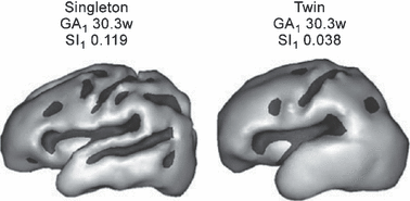
3D representation of the inner cortical surface for a singleton and a twin of equivalent age. SI1 represents the average of the sulcation index. Note the altered cortical gyrification shown to be significantly affected in twins when compared to singleton of the same gestational age. Modified from Dubois et al. (2008a).
Fig. 4
Fig. 4
(A) Cortical surface of ferret brains at 4, 10, 17 days of life and in an adult. (B) Cortical surface of human brains at 25, 30, 33, 39 weeks of gestation and in an adult. Courtesy of Barnettte et al. (Barnette et al., 2009).
Fig. 5
Fig. 5
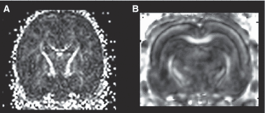
(A) Healthy preterm infant born at 29 weeks of gestation imaged at term equivalent, coronal FA map at the level of the posterior limb of the internal capsule. (B) P5 live rat pup, coronal FA map with sufficient in plane resolution to identify major white matter bundles such as the corpus callosum or the internal capsule. Image acquired at 11.7 Tesla in collaboration with Gregory Lodygensky and Jeffrey J Neil (Washington University, St Louis, Missouri).
Fig. 6
Fig. 6
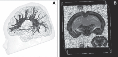
(A) 3D representation of the cortical surface of an infant at term with superimposed fiber tracking through the corpus callosum. Courtesy Jessica Dubois (CEA/SAC/DSV/DRM/NeuroSpin/Cognitive Neuroimaging Unit, Gif-sur-Yvette, France) (B) Relative anisotropy map of a fixed rat brain at 21 days of age with the superimposed fiber tracking through the corpus callosum. Image acquired at 9.4 Tesla in collaboration with Gregory Lodygensky and Rolf Gruetter (CIBM, Ecole polytechnique fédérale de Lausanne, Switzerland).
Fig. 7
Fig. 7
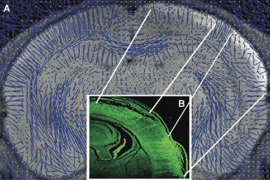
Principal eigenvector plot representing a disruption in the parietal cortex of the radially organized cortical eigenvectors 24 h after hypoxia-ischemia. (B) Fluoro-Jade B stain showing degenerating neurons in the same area. Courtesy of Sizonenko et al. (Sizonenko et al., 2007b).

References

    1. Abernethy LJ, Palaniappan M, Cooke RW. Quantitative magnetic resonance imaging of the brain in survivors of very low birth weight. Arch Dis Child. 2002;87:279–283. - PMC - PubMed
    1. Allin M, Matsumoto H, Santhouse AM, et al. Cognitive and motor function and the size of the cerebellum in adolescents born very pre-term 3. Brain. 2001;124:60–66. - PubMed
    1. Anbeek P, Vincken KL, Groenendaal F, et al. Probabilistic brain tissue segmentation in neonatal magnetic resonance imaging. Pediatr Res. 2008;63:158–163. - PubMed
    1. Anderson NG, Laurent I, Woodward LJ, et al. Detection of impaired growth of the corpus callosum in premature infants. Pediatrics. 2006;118:951–960. - PubMed
    1. Anjari M, Srinivasan L, Allsop JM, et al. Diffusion tensor imaging with tract-based spatial statistics reveals local white matter abnormalities in preterm infants. Neuroimage. 2007;35:1021–1027. - PubMed

MeSH terms