Comparative Study
. 2011 Feb;24(1):136-47.
doi: 10.1111/j.1755-148X.2010.00764.x. Epub 2010 Oct 6.

The deceptive nature of UVA tanning versus the modest protective effects of UVB tanning on human skin

Affiliations
Comparative Study

The deceptive nature of UVA tanning versus the modest protective effects of UVB tanning on human skin

Yoshinori Miyamura et al. Pigment Cell Melanoma Res. 2011 Feb.

Abstract

The relationship between human skin pigmentation and protection from ultraviolet (UV) radiation is an important element underlying differences in skin carcinogenesis rates. The association between UV damage and the risk of skin cancer is clear, yet a strategic balance in exposure to UV needs to be met. Dark skin is protected from UV-induced DNA damage significantly more than light skin owing to the constitutively higher pigmentation, but an as yet unresolved and important question is what photoprotective benefit, if any, is afforded by facultative pigmentation (i.e. a tan induced by UV exposure). To address that and to compare the effects of various wavelengths of UV, we repetitively exposed human skin to suberythemal doses of UVA and/or UVB over 2 weeks after which a challenge dose of UVA and UVB was given. Although visual skin pigmentation (tanning) elicited by different UV exposure protocols was similar, the melanin content and UV-protective effects against DNA damage in UVB-tanned skin (but not in UVA-tanned skin) were significantly higher. UVA-induced tans seem to result from the photooxidation of existing melanin and its precursors with some redistribution of pigment granules, while UVB stimulates melanocytes to up-regulate melanin synthesis and increases pigmentation coverage, effects that are synergistically stimulated in UVA and UVB-exposed skin. Thus, UVA tanning contributes essentially no photoprotection, although all types of UV-induced tanning result in DNA and cellular damage, which can eventually lead to photocarcinogenesis.


Figures

Figure 1
Figure 1
Increased melanin content and CPD damage of pigmented human skin equivalents after UV challenge. (Top) Images of melanin content in human skin equivalents from White, Asian, and Black were analyzed by Fontana-Masson staining and data are reported as mean density; scale bar = 50 μm. (Bottom) The unexposed control, or skin equivalents exposed to a UV challenge of 50 mJ/cm2 were harvested at day 0 (left column) and at day 9 (right column) immediately after the UV challenge. TDM2 was used as the primary antibody to detect CPD and is stained green; scale bar = 50 μm. Images were analyzed as noted in the Methods, and CPD levels are expressed as % CPD-positive area (mean ± SEM, n=10). Statistical significance of differences from the day 0 control are indicated (NS = not significant).
Figure 2
Figure 2
Tanning responses in the skin following UV exposure. (Left) Representative image of tanning responses induced in subject 2 after 2 weeks of suberythemal repetitive exposure to UVA & UVB. (Right) Vector representation of the UVA&UVB exposed skin versus the unexposed control in the L* and b* plane of the CIE L*a*b* color space system. These parameters allow for calculation of the individual typology angle (ITA°) as defined in the Methods and allow for segmentation into several pigmentation categories.
Figure 3
Figure 3
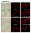
Melanin content and CPD damage in the skin following UV exposure. (Left) Representative sections from subject 5 were stained using the Fontana-Masson method to evaluate melanin content. (Center) Unexposed (control), UVA-, UVB- and UVA&UVB-exposed skin specimens as noted were biopsied before or after (Right) the 2 MED UV challenge; images from subject 5 are shown as an example. TDM2 was used as the primary antibody to detect CPD and is stained red due to the emission of the Alexa 568 conjugated secondary antibody; scale bar = 50 μm. Images were analyzed as noted in the Methods, and CPD levels are expressed as % CPD-positive area (mean ± SEM, n=7). CPD levels are expressed as mean fluorescence intensity as follows: Control – No Challenge 0.19 ± 0.05, Control – UV Challenge 0.87 ± 0.10, p < 0.0005; UVA&UVB – No Challenge 0.52 ± 0.09, UVA&UVB – UV Challenge 0.68 ± 0.09, p < 0.05; UVA – No Challenge 0.29 ± 0.04, UVA – UV Challenge 0.78 ± 0.09, p < 0.002; UVB – No Challenge 0.51 ± 0.06, UVB – UV Challenge 0.80 ± 0.06, p < 0.01. In addition CPD statistical significance of differences from the corresponding unchallenged areas is indicated.
Figure 4
Figure 4
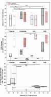
Relationship of UV exposure to CPD damage and melanin content. (Top) Interindividual distribution of % melanin positive area and (Middle) CPD positive area graphed as box plots for unexposed (control), UVA-, UVB- and UVA&UVB-exposed skin as well as the areas receiving the 2 MED UV challenge denoted with a red boundary (the dashed line in each box plot represents the mean, the solid line the median value). (Bottom) The % change in DNA damage after the 2 MED UV challenge was calculated and shows an inverse correlation relative to the mean % melanin positive area. * p < 0.05, ** p < 0.01, *** p < 0.005, **** p < 0.0005, not significant comparisons are absent from graphs.
Figure 5
Figure 5
Relationship of UV tanning to the distribution of melanin. (Top) High magnification light microscopy images of representative areas of skin stained by the Fontana Masson method; example of pigment selection defined as stratum basale layer = red, stratum spinosum layer = green, stratum granulosum layer = blue, total epidermis = cyan to determine pigment granule size in μm2; scale bar = 20 μm. (Middle) Images were captured and analyzed as described in the Methods, and data are reported as box plots of size distribution of pigment granules in the 3 epidermal layers as well as in the total epidermis within 100X images (n=6 subjects, the solid line shows the median value). (Bottom) Electron micrographs taken of specimens from subject 8; scale bar = 1 μm.

Similar articles

Cited by

References

    1. Abdel-Malek ZA, Kadekaro AL, Swope VB. Stepping up melanocytes to the challenge of UV exposure. Pigment Cell Melanoma Res. 2010;23:171–186. - PubMed
    1. Agar N, Young AR. Melanogenesis: a photoprotective response to DNA damage? Mutat. Res. 2005;571:121–132. - PubMed
    1. Agar NS, Halliday GM, Barnetson RS, Ananthaswamy HN, Wheeler M, Jones AM. The basal layer in human squamous tumors harbors more UVA than UVB fingerprint mutations: a role for UVA in human skin carcinogenesis. Proc. Natl. Acad. Sci. USA. 2004;101:4954–4959. - PMC - PubMed
    1. Bennett DC. Ultraviolet wavebands and melanoma initiation. Pigment Cell Melanoma Res. 2008;21:520–524. - PubMed
    1. Berneburg M, Grether-Beck S, Kurten V, Ruzicka T, Briviba K, Sies H, Krutmann J. Singlet oxygen mediates the UVA-induced generation of the photoaging-associated mitochondrial common deletion. J. Biol. Chem. 1999;274:15345–15349. - PubMed

Publication types