Review
. 2010 Nov;51(11):1821-39.
doi: 10.1093/pcp/pcq156. Epub 2010 Oct 26.

Molecular basis of the core regulatory network in ABA responses: sensing, signaling and transport

Affiliations
Review

Molecular basis of the core regulatory network in ABA responses: sensing, signaling and transport

Taishi Umezawa et al. Plant Cell Physiol. 2010 Nov.

Abstract

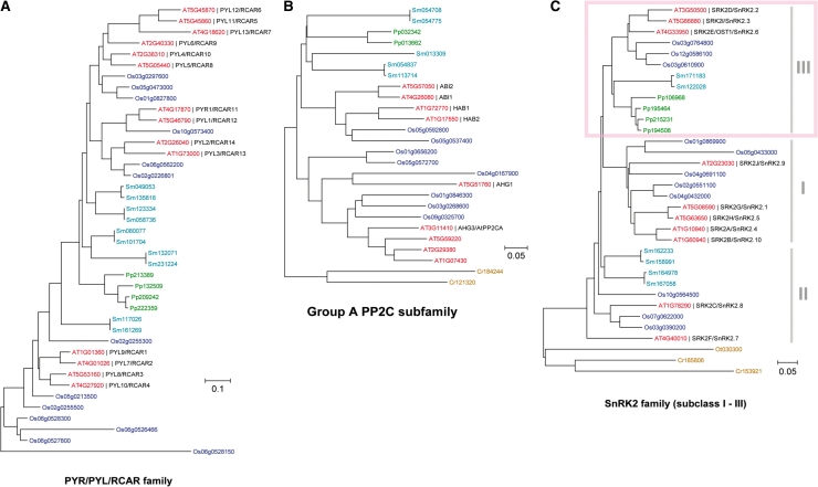
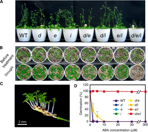
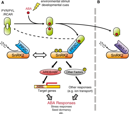
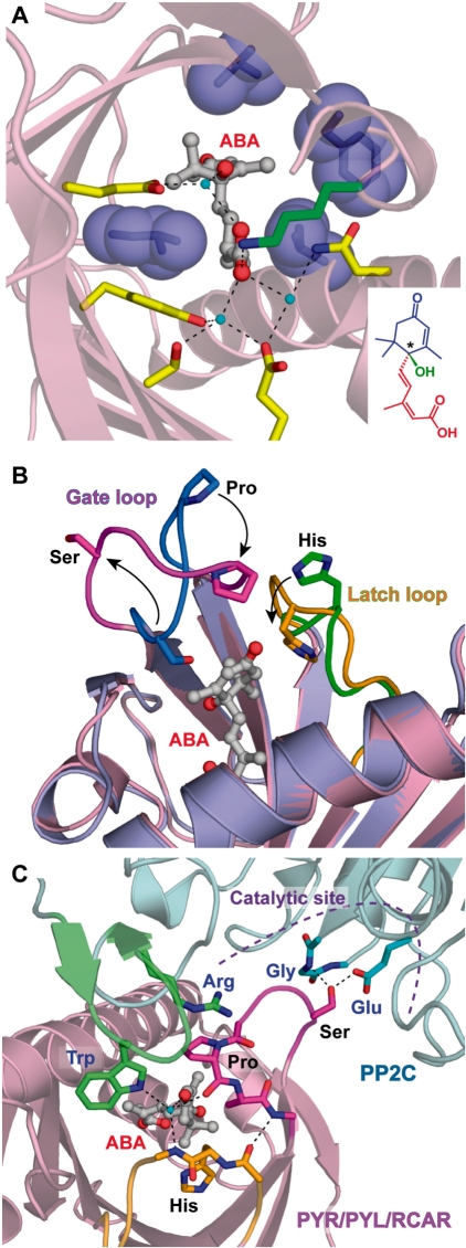
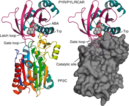
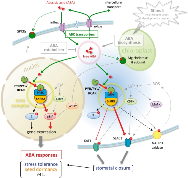
ABA is a major phytohormone that regulates a broad range of plant traits and is especially important for adaptation to environmental conditions. Our understanding of the molecular basis of ABA responses in plants improved dramatically in 2009 and 2010, banner years for ABA research. There are three major components; PYR/PYL/ RCAR (an ABA receptor), type 2C protein phosphatase (PP2C; a negative regulator) and SNF1-related protein kinase 2 (SnRK2; a positive regulator), and they offer a double negative regulatory system, [PYR/PYL/RCAR-| PP2C-| SnRK2]. In the absence of ABA, PP2C inactivates SnRK2 by direct dephosphorylation. In response to environmental or developmental cues, ABA promotes the interaction of PYR/PYL/RCAR and PP2C, resulting in PP2C inhibition and SnRK2 activation. This signaling complex can work in both the nucleus and cytosol, as it has been shown that SnRK2 phosphorylates basic-domain leucine zipper (bZIP) transcription factors or membrane proteins. Several structural analyses of PYR/PYL/RCAR have provided the mechanistic basis for this 'core signaling' model, by elucidating the mechanism of ABA binding of receptors, or the 'gate-latch-lock' mechanism of interaction with PP2C in inhibiting activity. On the other hand, intercellular ABA transport had remained a major issue, as had intracellular ABA signaling. Recently, two plasma membrane-type ABC transporters were identified and shed light on the influx/efflux system of ABA, resolving how ABA is transported from cell to cell in plants. Our knowledge of ABA responses in plants has been greatly expanded from intracellular signaling to intercellular transport of ABA.


Figures

Fig. 1
Fig. 1
Phylogenetic trees of the core components in ABA signaling. Phylogenetic trees of PYR/PYL/RCAR (A), group A PP2C (B) and the SnRK2 family (C) from several plant species. Amino acid sequences for each family were retrieved from Arabidopsis (red), rice (blue), Selaginella moellendorffii (azure blue), Physcomitrella patens (green), Ostreococcus tauri and Chlamydomonas reinhardtii (yellow ocher) using the SALAD database (http://salad.dna.affrc.go.jp/CGViewer/). The tree was drawn using the Neighbor–Joining method in the MEGA 4.0 program.
Fig. 2
Fig. 2
Subclass III SnRK2 protein kinases are essential for the control of drought tolerance and germination. (A) The srk2dei (snrk2.2 snrk2.3 snrk2.6) triple mutants exhibited greatly reduced tolerance to drought stress. (B) Viviparous seeds in attached siliques of an srk2dei mutant grown in high humidity. (C) Extreme ABA insensitivity of srk2dei mutant plants. These figures are modified from Nakashima et al. (2009) and Fujita et al. (2009).
Fig. 3
Fig. 3
Proposed model of the major ABA signaling pathway. PYR/PYL/RCAR, PP2C and SnRK2 form a signaling complex referred to as the ‘ABA signalosome’. (A) Under normal conditions, PP2C negatively regulates SnRK2 by direct interactions and dephosphorylation of multiple residues of SnRK2. Once abiotic stresses or developmental cues up-regulate endogenous ABA, PYR/PYL/RCAR binds ABA and interacts with PP2C to inhibit protein phosphatase activity. In turn, SnRK2 is released from PP2C-dependent regulation and activated to phosphorylate downstream factors, such as the AREB/ABF bZIP-type transcription factor or membrane proteins involving ion channels. (B) In contrast, the abi1-1-type mutated protein lacks PYR/PYL/RCAR binding, resulting in the constitutive inactivation of SnRK2, even in the presence of ABA, and strong insensitivity to ABA in the abi1-1 mutant.
Fig. 4
Fig. 4
Structural analysis of PYR/PYL/RCAR ABA receptors. (A) Stereoselective ABA-binding mode of the PYR/PYL/RCAR proteins. A lysine residue (green) directly interacts with ABA, and polar residues (yellow) form a water-mediated hydrogen bond network with ABA. Water molecules and hydrogen bonds are shown by cyan spheres and dashed lines, respectively. Hydrophobic residues (blue) are localized around dimethyl and monomethyl groups of the cyclohexene moiety. For the structural formula of ABA in the inset, the cyclohexene moiety, pentadienoic acid moiety and hydroxyl group from the chiral carbon (shown by an asterisk) are colored blue, red and green, respectively. This figure was created using Protein Data Bank (PDB) coordinates of ABA-bound PYL1 (3JRS). (B) Open-to-closed gating mechanism of the PYR/PYL/RCAR proteins. Gate and latch loops dramatically shift to the closed conformation (magenta and orange, respectively) from the open conformation (blue and green, respectively) upon ABA binding. This figure was created using the PDB coordinates of apo-PYL1 (3KAY) and ABA-bound PYL1 (3JRS). (C) ABA-dependent mechanism for PP2C inhibition. Conserved tryptophan and arginine residues on the additional antiparallel β-sheet of PP2C (green) contact the gate and latch loops of the PYR/PYL/RCAR proteins. The gate loop is locked by these interactions and seals the catalytic site of PP2C to inhibit phosphatase activity competitively. This figure was created using PDB coordinates from apo-PYL1 (3KAY), ABA-bound PYL1 (3JRS) and the complex of ABA-bound PYL1 and ABI1 (3JRQ).
Fig. 5
Fig. 5
Overall structure of the complex of ABA-bound PYL1 and PP2C. PP2C in the left and right diagrams is represented by a ribbon and surface model, respectively. This figure was created using PDB coordinates for the complex of ABA-bound PYL1 and ABI1 (3JRQ).
Fig. 6
Fig. 6
Evolution of core components of ABA signaling. As shown in Fig. 1, PYR/PYL/RCAR, group A PP2C and subclass III SnRK2 are conserved from bryophytes. The development of an ABA signaling system seems to be highly correlated with the evolution from aquatic to terrestrial plants. As representatives, component numbers of bryophyte, lycophyte and angiosperm were obtained from Physcomitrella patens, Selaginella moellendorffii and Arabidopsis thaliana, respectively.
Fig. 7
Fig. 7
Schematic view of hypothetical ABA intercellular transmission. This diagram is an Arabidopsis leaf section showing two distinct cell types: vascular tissues, including vascular parenchyma cells, and guard cells on the leaf epidermis. AtABCG25 might function in ABA efflux from ABA-biosynthesizing vascular cells, and ABA would diffuse into apoplastic areas. AtABCG40 might function in ABA influx into guard cells to facilitate stomatal closure.
Fig. 8
Fig. 8
Overview of ABA sensing, signaling and transport. The ABA-related components described in this review are summarized. PYR/PYL/RCAR, PP2C and SnRK2 form a core signaling complex (yellow circle), which functions in at least two sites. One is the nucleus, in which the core complex directly regulates ABA-responsive gene expression by phosphorylation of AREB/ABF-type transcription factors. The other is the cytoplasm, and the core complex can access the plasma membrane and phosphorylate anion channels (SLAC1) or potassium channels (KAT1) to induce stomatal closure in response to ABA. Other substrates of SnRK2s have yet to be identified. The principal mechanism of ABA sensing or signaling in the core signaling complex is illustrated in Fig. 2. In contrast, the endogenous ABA level is a major determinant of ABA sensing that is maintained by ABA biosynthesis, catabolism or transport. The ABA transport system consists of two types of ABC transporter for influx or efflux. Although ABA biosynthesis and catabolism were not the focus of this review, these types of regulation are well described in other reviews (e.g. Nambara and Marion-Poll 2005, Hirayama and Shinozaki 2010). ABA movements are indicated by green lines and arrows, and major signaling pathways are indicated by red lines and arrows. Dotted lines indicate indirect or unconfirmed connections.

References

    1. Anderberg RJ, Walker-Simmons MK. Isolation of a wheat cDNA clone for an abscisic acid-inducible transcript with homology to protein kinases. Proc. Natl Acad. Sci. USA. 1992;89:10183–10187. - PMC - PubMed
    1. Belin C, de Franco P, Bourbousse C, Chaignepain S, Schmitter J, Vavasseur A, et al. Identification of features regulating OST1 kinase activity and OST1 function in guard cells. Plant Physiol. 2006;141:1316–1327. - PMC - PubMed
    1. Bensmihen S, Giraudat J, Parcy F. Characterization of three homologous basic leucine zipper transcription factors (bZIP) of the ABI5 family during Arabidopsis thaliana embryo maturation. J. Exp. Bot. 2005;56:597–603. - PubMed
    1. Bensmihen S, Rippa S, Lambert G, Jublot D, Pautot V, Granier F, et al. The homologous ABI5 and EEL transcription factors function antagonistically to fine-tune gene expression during late embryogenesis. Plant Cell. 2002;14:1391–1403. - PMC - PubMed
    1. Boudsocq M, Barbier-Brygoo H, Lauriere C. Identification of nine sucrose nonfermenting 1-related protein kinases 2 activated by hyperosmotic and saline stresses in Arabidopsis thaliana. J. Biol. Chem. 2004;279:41758–41766. - PubMed

Publication types