

High connectivity between reduced cortical thickness and disrupted white matter tracts in long-standing type 1 diabetes
- PMID: 20980455
- PMCID: PMC3012188
- DOI: 10.2337/db10-0598
High connectivity between reduced cortical thickness and disrupted white matter tracts in long-standing type 1 diabetes
Abstract
Objective: Previous studies have observed disruptions in brain white and gray matter structure in individuals with type 1 diabetes, and these structural differences have been associated with neurocognitive testing deficiencies. This study investigated the relationship between cerebral cortical thickness reductions and white matter microstructural integrity loss in a group of patients with type 1 diabetes and in healthy control subjects using diffusion tensor imaging (DTI).
Research design and methods: Twenty-five subjects with type 1 diabetes for at least 15 years and 25 age- and sex-matched control subjects underwent structural T1 and proton-density and DTI on a 3.0 Tesla scanner. Fractional anisotropy measurements were made on major cerebral white matter tracts, and DTI tractography was performed to identify cortical regions with high connectivity to these tracts.
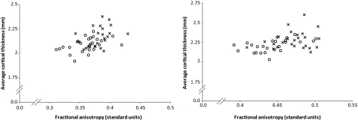
Results: Posterior white matter tracts with reduced fractional anisotropy (optic radiations, posterior corona radiata, and the splenium region of the corpus callosum) were found to have high connectivity with a number of posterior cortical regions, including the cuneus, precuneus, fusiform, and posterior parietal cortical regions. A significant reduction in cortical thickness in the diabetic group was observed in the regions with high connectivity to the optic radiations and posterior corona radiata tracts (P < 0.05).
Conclusions: The direct relationship between white and gray matter structural pathology has not been previously demonstrated in subjects with long-standing type 1 diabetes. The relationship between posterior white matter microstructural integrity disruption and lower cortical thickness demonstrated using a novel DTI connectivity technique suggests a common or interrelated pathophysiological mechanism in type 1 diabetes.
Figures


References
-
- Howorka K, Pumprla J, Saletu B, Anderer P, Krieger M, Schabmann A: Decrease of vigilance assessed by EEG-mapping in type I diabetic patients with history of recurrent severe hypoglycaemia. Psychoneuroendocrinology 2000;25:85–105 - PubMed
-
- Pozzessere G, Rizzo PA, Valle E, Mollica MA, Meccia A, Morano S, Di Mario U, Andreani D, Morocutti C: Early detection of neurological involvement in IDDM and NIDDM: multimodal evoked potentials versus metabolic control. Diabetes Care 1988;11:473–480 - PubMed
Publication types
MeSH terms
Grants and funding
LinkOut - more resources
Full Text Sources
Medical
