. 2011 Feb;60(2):590-601.
doi: 10.2337/db10-0403. Epub 2010 Oct 27.

The protective role of Smad7 in diabetic kidney disease: mechanism and therapeutic potential

Affiliations

The protective role of Smad7 in diabetic kidney disease: mechanism and therapeutic potential

Hai Yong Chen et al. Diabetes. 2011 Feb.

Abstract

Objective: Although Smad3 has been considered as a downstream mediator of transforming growth factor-β (TGF-β) signaling in diabetes complications, the role of Smad7 in diabetes remains largely unclear. The current study tests the hypothesis that Smad7 may play a protective role and has therapeutic potential for diabetic kidney disease.

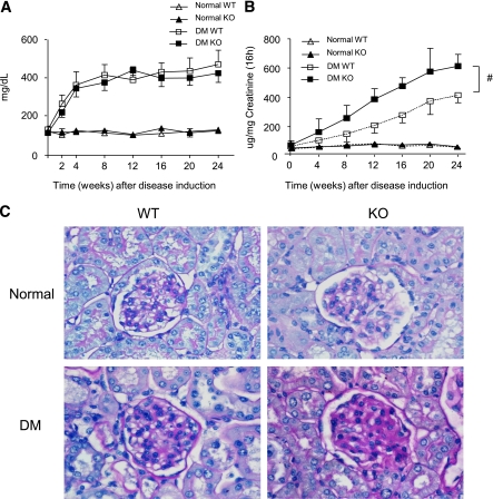
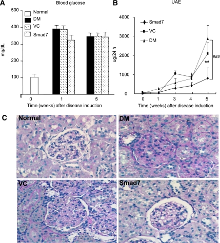
Research design and methods: Protective role of Smad7 in diabetic kidney disease was examined in streptozotocin-induced diabetic mice that have Smad7 gene knockout (KO) and in diabetic rats given Smad7 gene transfer using an ultrasound-microbubble-mediated technique.

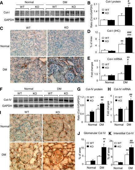
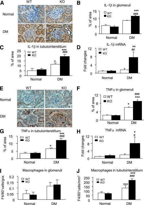
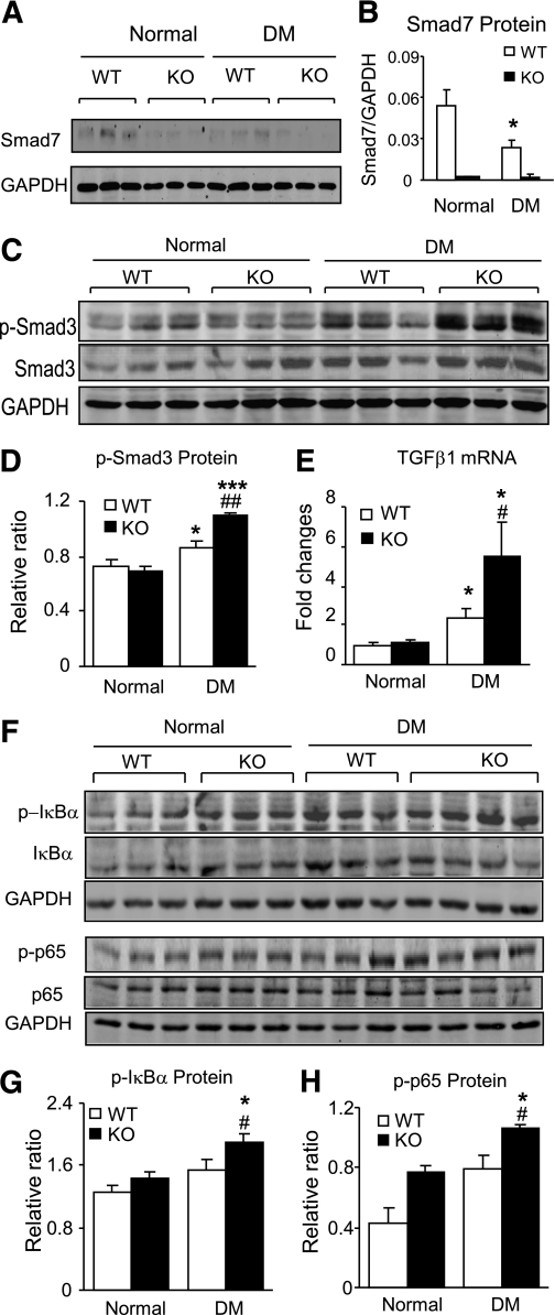
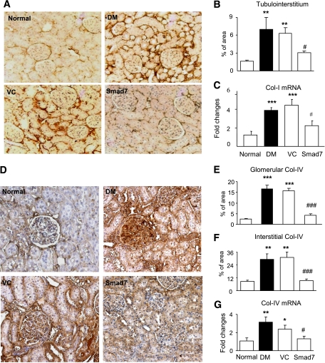
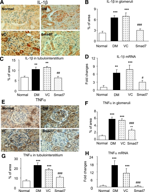
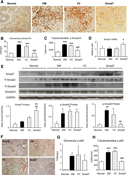
Results: We found that mice deficient for Smad7 developed more severe diabetic kidney injury than wild-type mice as evidenced by a significant increase in microalbuminuria, renal fibrosis (collagen I, IV, and fibronectin), and renal inflammation (interleukin-1β [IL-1β], tumor necrosis factor-α [TNF-α], monocyte chemoattractant protein-1 [MCP-1], intracellular adhesion molecule-1 [ICAM-1], and macrophages). Further studies revealed that enhanced renal fibrosis and inflammation in Smad7 KO mice with diabetes were associated with increased activation of both TGF-β/Smad2/3 and nuclear factor-κB (NF-κB) signaling pathways. To develop a therapeutic potential for diabetic kidney disease, Smad7 gene was transferred into the kidney in diabetic rats by an ultrasound-microbubble-mediated technique. Although overexpression of renal Smad7 had no effect on levels of blood glucose, it significantly attenuated the development of microalbuminuria, TGF-β/Smad3-mediated renal fibrosis such as collagen I and IV and fibronectin accumulation and NF-κB/p65-driven renal inflammation including IL-1β, TNF-α, MCP-1, and ICAM-1 expression and macrophage infiltration in diabetic rats.

Conclusions: Smad7 plays a protective role in diabetic renal injury. Overexpression of Smad7 may represent a novel therapy for the diabetic kidney complication.


Figures

FIG. 1.
FIG. 1.
Diabetic kidney injury is enhanced in Smad7 KO mice. A: Blood glucose. Levels of blood glucose are significantly increased at week 2 after STZ injection and maintained at equal higher levels over the 24-week period in both Smad7 KO and WT mice. B: Urinary albumin exertion (UAE). Smad7 KO mice had more severe microalbuminuria than the WT mice. C: Histology (PAS-stained sections). Data are expressed as mean ± SE for group of eight mice. #P < 0.05 vs. WT DM group by 2-way ANOVA. Magnification ×400. (A high-quality color representation of this figure is available in the online issue.)
FIG. 2.
FIG. 2.
Renal fibrosis is enhanced in diabetic Smad7 KO mice. AE: Collagen I (Col I) expression. FK: Collagen IV (Col IV) expression. Results show that when compared with the WT mice, Smad7 KO mice significantly enhance renal collagen I and IV expression as demonstrated by Western blot analysis (A, B, F, and G), immunohistochemistry (IHC; C and I) and quantitative analysis in glomeruli (J) and tubulointerstitium (D and K), and real-time PCR at the mRNA level (E and H). Data represent mean ± SE for groups of eight animals. *P < 0.05, **P < 0.01, ***P < 0.001 vs. normal; #P < 0.05, ##P < 0.01, ###P < 0.001 vs. WT DM mice. Magnification ×400 (A and F). (A high-quality color representation of this figure is available in the online issue.)
FIG. 3.
FIG. 3.
Renal inflammation is enhanced in diabetic Smad7 KO mice. AD: IL-1β expression. EH: TNF-α expression. I and J: Macrophages infiltrating the glomerulus and the tubulointerstitium. Results show that when compared with the WT mice, Smad7 KO mice develop more severe renal inflammation by enhancing renal IL-1β and TNF-α expression in glomeruli (A, B, E, and F) and in renal cortex (C and G) as demonstrated by immunohistochemistry and mRNA expression real-time PCR (D and H). Data represent mean ± SE for groups of eight animals. *P < 0.05, **P < 0.01, ***P < 0.001 vs. normal; #P < 0.05, ##P < 0.01, ###P < 0.001 vs. WT DM mice. Magnification ×400 (A and E). (A high-quality color representation of this figure is available in the online issue.)
FIG. 4.
FIG. 4.
Enhanced TGF-β/Smad and NF-κB signaling pathways in diabetic Smad7 KO mice. Western blot analysis (A and B) shows that when compared with normal WT mice, expression of Smad7 is significantly decreased in diabetic WT mice but undetectable in both normal and diabetic KO mice. C and D: Phosphorylation of Smad3. Western blot analysis shows an enhanced TGF-β/Smad3 signaling (p-Smad3) in diabetic Smad7 mice compared with diabetic WT mice. E: Real-time PCR detects an increased renal TGF-β1 mRNA expression in diabetic Smad7 KO mice. F: Activation of NF-κB. Western blot analysis reveals that an increased phosphorylation of IκBα in Smad7 KO mice is associated with enhanced phosphorylation of NF-κB/p65 (p-p65). Note that phosphorylation causes degradation of IκBα and NF-κB/p65 protein, respectively. J: Quantitation of phosphorylated IκBα (p-IκBα). H: Quantitation of phosphorylated p65 (p-p65). Data represent mean ± SE for groups of eight animals. *P < 0.05, ***P < 0.001 vs. normal; #P < 0.05, ##P < 0.01 vs. WT DM mice.
FIG. 5.
FIG. 5.
Smad7 gene therapy attenuates diabetic kidney injury in rats. A: Blood glucose. B: Microalbuminuria. C: Histology. When compared with the diabetic rats (DM) and empty vector control DM rats (VC), Smad7 gene therapy attenuates microalbuminuria and ECM deposition within the glomerulus and tubulointerstitium. Data represent mean ± SE for groups of six animals. **P < 0.01 vs. normal; ###P < 0.001 vs. DM and VC. (A high-quality color representation of this figure is available in the online issue.)
FIG. 6.
FIG. 6.
Overexpression of renal Smad7 inhibits renal fibrosis in diabetic rats. AC: Collagen I expression. DG: Collagen IV expression. Results show that when compared with diabetic rats (DM) and diabetic rats with control vector treatment (VC), Smad7 gene transfer significantly inhibits renal collagen I and IV expression as demonstrated by immunohistochemistry (A, B, and DF) and real-time PCR at the mRNA levels (C and G). Data represent mean ± SE for groups of six animals. *P < 0.05, **P < 0.01, ***P < 0.001 vs. normal; #P < 0.05, ###P < 0.001 vs. DM and VC mice. Magnification ×200 (A and D). (A high-quality color representation of this figure is available in the online issue.)
FIG. 7.
FIG. 7.
Overexpression of renal Smad7 inhibits renal inflammation in diabetic rats. AD: IL-1β expression. EH: TNF-α expression. Immunohistochemistry and real-time PCR demonstrate that when compared with diabetic rats (DM) and diabetic rats treated with control vector (VC), Smad7 gene transfer significantly inhibits renal inflammation including IL-1β and TNF-α protein expression in glomeruli (A, B, E, and F) and tubulointerstitium (C and G) and their mRNA expression (D and H). Data represent mean ± SE for groups of six animals. *P < 0.05, **P < 0.01, ***P < 0.001 vs. normal; #P < 0.05, ##P < 0.01, ###P < 0.001 vs. DM and VC mice. Magnification ×400 (A and E). (A high-quality color representation of this figure is available in the online issue.)
FIG. 8.
FIG. 8.
Overexpression of renal Smad7 blocks activation of the TGF-β/Smad and NF-κB signaling pathways in diabetic rats. A: Phosphorylated Smad2/3 (p-Smad2/3) nuclear location. B and C: Quantitative analysis of nucleated p-Smad2/3 in glomeruli and tubulointerstitium. D: Renal Smad7 mRNA expression by real-time PCR. E: Western blot analysis of renal Smad7 and phosphorylation of Smad2 (p-Smad2) and Smad3 (p-Smad3). Note that Smad7 gene transfer upregulates renal Smad7, thereby inhibiting phosphorylation of Smad3, but not Smad2. F: Phosphorylated NF-κB/p65 nuclear location. G and H: Quantitative analysis of phosphorylated NF-κB/p65 in the glomerulus and tubulointerstitium. Data represent mean ± SE for groups of six animals. *P < 0.05, **P < 0.01, ***P < 0.001 vs. normal; ##P < 0.05, ###P < 0.001 vs. DM and VC mice. Magnification ×400 (A and F). (A high-quality color representation of this figure is available in the online issue.)

References

    1. Caramori ML, Kim Y, Huang C, et al. Cellular basis of diabetic nephropathy: 1. Study design and renal structural-functional relationships in patients with long-standing type 1 diabetes. Diabetes 2002;51:506–513 - PubMed
    1. Mauer SM, Steffes MW, Ellis EN, Sutherland DE, Brown DM, Goetz FC. Structural-functional relationships in diabetic nephropathy. J Clin Invest 1984;74:1143–1155 - PMC - PubMed
    1. Pagtalunan ME, Miller PL, Jumping-Eagle S, et al. Podocyte loss and progressive glomerular injury in type II diabetes. J Clin Invest 1997;99:342–348 - PMC - PubMed
    1. White KE, Bilous RW. Type 2 diabetic patients with nephropathy show structural-functional relationships that are similar to type 1 disease. J Am Soc Nephrol 2000;11:1667–1673 - PubMed
    1. Finne P, Reunanen A, Stenman S, Groop PH, Grönhagen-Riska C. Incidence of end-stage renal disease in patients with type 1 diabetes. JAMA 2005;294:1782–1787 - PubMed

Publication types

MeSH terms