. 2011 Jan;85(1):378-89.
doi: 10.1128/JVI.01900-10. Epub 2010 Oct 27.

Cytomegalovirus microRNA expression is tissue specific and is associated with persistence

Affiliations

Cytomegalovirus microRNA expression is tissue specific and is associated with persistence

Christine Meyer et al. J Virol. 2011 Jan.

Abstract

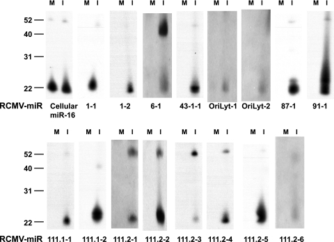
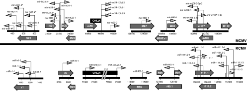
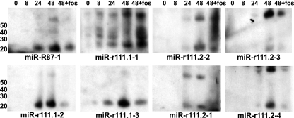
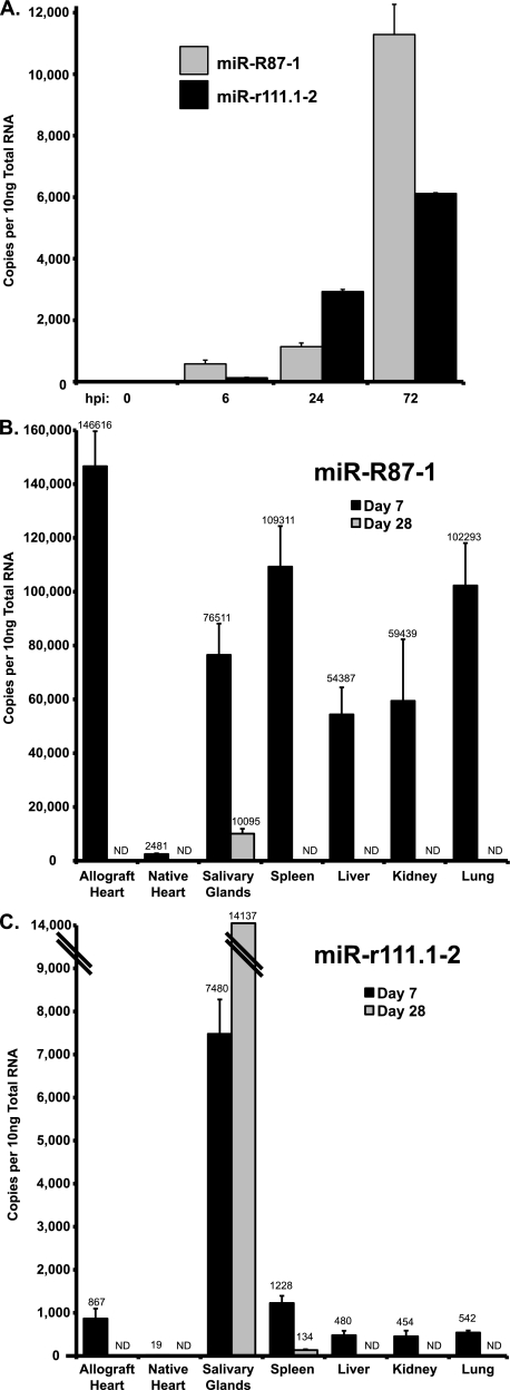
MicroRNAs (miRNAs) are a class of small noncoding RNAs involved in posttranscriptional regulation. miRNAs are utilized in organisms ranging from plants to higher mammals, and data have shown that DNA viruses also use this method for host and viral gene regulation. Here, we report the sequencing of the small RNAs in rat cytomegalovirus (RCMV)-infected fibroblasts and persistently infected salivary glands. We identified 24 unique miRNAs that mapped to hairpin structures found within the viral genome. While most miRNAs were detected in both samples, four were detected exclusively in the infected fibroblasts and two were specific for the infected salivary glands. The RCMV miRNAs are distributed across the viral genome on both the positive and negative strands, with clusters of miRNAs at a number of locations, including near viral genes r1 and r111. The RCMV miRNAs have a genomic positional orientation similar to that of the miRNAs described for mouse cytomegalovirus, but they do not share any substantial sequence conservation. Similar to other reported miRNAs, the RCMV miRNAs had considerable variation at their 3' and 5' ends. Interestingly, we found a number of specific examples of differential isoform usage between the fibroblast and salivary gland samples. We determined by real-time PCR that expression of the RCMV miRNA miR-r111.1-2 is highly expressed in the salivary glands and that miR-R87-1 is expressed in most tissues during the acute infection phase. Our study identified the miRNAs expressed by RCMV in vitro and in vivo and demonstrated that expression is tissue specific and associated with a stage of viral infection.


Figures

FIG. 1.
FIG. 1.
Northern blot analysis of RCMV miRNAs. RFL6 cells were infected at a multiplicity of 1 PFU per cell and subjected to Northern blot analysis using probes specific for the predicted RCMV miRNA sequences. Lanes M, mock infected; lanes I, infected. The cellular miRNA miR-16 was used as a loading control. The values to the left are molecular sizes in nucleotides.
FIG. 2.
FIG. 2.
Genomic organization of RCMV miRNAs. The diagram depicts the regions of the RCMV genome that contain RCMV miRNAs. The dark gray arrows indicate the positions and directions of the RCMV miRNAs, where right-pointing arrows indicate RCMV miRNAs on the sense strand and left-pointing arrows indicate miRNAs on the complement strand. Open reading frames that contain or are near viral miRNAs are shown as light gray block arrows. The RCMV origin of lytic replication is depicted as a dark gray box.
FIG. 3.
FIG. 3.
Secondary structures of RCMV precursor miRNAs. The predicted stem-loop secondary structures of the RCMV pre-miRNAs from the Mfold program (68) are depicted with the mature miRNAs shaded in gray. When two miRNAs are transcribed from one stem-loop, the position of each miRNA is indicated on the 5′ or 3′ arm of the stem-loop.
FIG. 4.
FIG. 4.
Comparison of genomic positioning of the RCMV and MCMV miRNAs. The diagram depicts the genomic positioning of the miRNAs expressed by RCMV (lower) and MCMV (upper). The dark gray arrows indicate the positions and directions of the CMV miRNAs, where right-pointing arrows indicate miRNAs on the sense strands and left-pointing arrows indicate miRNAs on the complement strands. Open reading frames that contain or are near viral miRNAs are shown as light gray block arrows. The MCMV and RCMV origins of lytic replication are depicted as black boxes.
FIG. 5.
FIG. 5.
Kinetic analysis of miRNA expression by Northern blotting. Total RNA was harvested with Trizol from RCMV-infected RFL6 rat fibroblasts at 0, 8, 24, and 48 hpi. Cells were infected at a MOI of 1.0. An additional sample harvested at 48 hpi was treated with Foscavir (foscarnet sodium; 100 mg/ml) to prevent late gene expression. RNA was subjected to Northern blot analysis using probes specific for the predicted RCMV miRNA sequences. Lane 1 (0 hpi) was a mock-infected sample. The values to the left are molecular sizes in nucleotides.
FIG. 6.
FIG. 6.
RCMV miRNA expression in tissues from RCMV-infected allograft recipients. The level of RCMV miRNA expression was determined by RT-PCR analysis of total RNA samples. Shown are viral miRNA copy numbers determined using dilutions of an oligonucleotide standard with the specific miRNA sequences of miR-R87-1 and miR-r111.1-2. (A) Quantification of RCMV miRNA miR-R87-1 and miR-r111.1-2 expression in RCMV-infected fibroblasts at 0, 6, 24, and 72 hpi. Uninfected cells (time zero) were included as a negative control. Both viral miRNAs accumulate with increasing time. (B) RT-PCR results for RCMV miR-R87-1 from tissues harvested from RCMV-infected heart allograft recipients (n = 3) at 7 and 28 days posttransplantation. RCMV miR-R87-1 was most highly expressed in the allograft heart, spleen, and lung tissues at 7 days posttransplantation. (C) RT-PCR results for RCMV miR-r111.1-2 from tissues harvested from RCMV-infected heart allograft recipients (n = 3). miR-r111.1-2 was most highly expressed in salivary glands at 7 and 28 days posttransplantation. ND, not detected.

References

    1. Ameres, S. L., M. D. Horwich, J. H. Hung, J. Xu, M. Ghildiyal, Z. Weng, and P. D. Zamore. 2010. Target RNA-directed trimming and tailing of small silencing RNAs. Science 328:1534-1539. - PMC - PubMed
    1. Babiarz, J. E., J. G. Ruby, Y. Wang, D. P. Bartel, and R. Blelloch. 2008. Mouse ES cells express endogenous shRNAs, siRNAs, and other Microprocessor-independent, Dicer-dependent small RNAs. Genes Dev. 22:2773-2785. - PMC - PubMed
    1. Bartel, D. P. 2004. MicroRNAs: genomics, biogenesis, mechanism, and function. Cell 116:281-297. - PubMed
    1. Bartel, D. P., and C. Z. Chen. 2004. Micromanagers of gene expression: the potentially widespread influence of metazoan microRNAs. Nat. Rev. Genet. 5:396-400. - PubMed
    1. Beisser, P. S., C. Vink, J. G. Van Dam, G. Grauls, S. J. Vanherle, and C. A. Bruggeman. 1998. The R33 G protein-coupled receptor gene of rat cytomegalovirus plays an essential role in the pathogenesis of viral infection. J. Virol. 72:2352-2363. - PMC - PubMed

Publication types