. 2011 Jan;85(1):146-55.
doi: 10.1128/JVI.01265-10. Epub 2010 Oct 27.

Adenovirus membrane penetration activates the NLRP3 inflammasome

Affiliations

Adenovirus membrane penetration activates the NLRP3 inflammasome

A U Barlan et al. J Virol. 2011 Jan.

Abstract

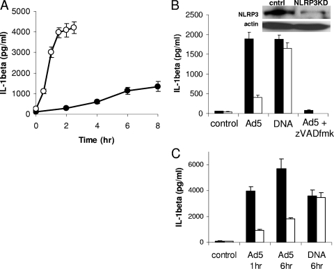
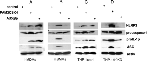
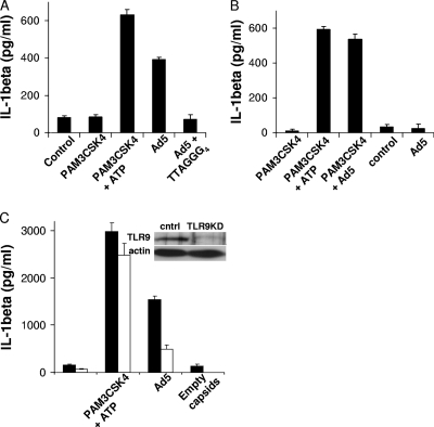
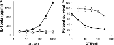
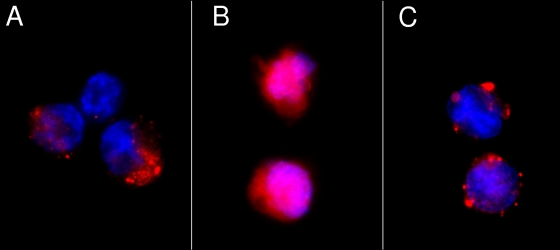
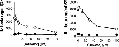
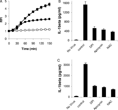
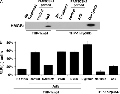
Adenovirus type 5 (Ad5) infection of macrophages results in rapid secretion of interleukin-1β (IL-1β) and is dependent on the inflammasome components NLRP3 and ASC and the catalytic activity of caspase-1. Using lentivirus-expressed short hairpin RNA (shRNA) and competitive inhibitors, we show that Ad-induced IL-1β release is dependent upon Toll-like receptor 9 (TLR9) sensing of the Ad5 double-stranded DNA (dsDNA) genome in human cell lines and primary monocyte-derived macrophages but not in mouse macrophages. Additionally, a temperature-sensitive mutant of Ad5 unable to penetrate endosomal membranes, ts1, is unable to induce IL-1β release in TLR2-primed THP-1 cells, suggesting that penetration of endosomal membranes is required for IL-1β release. Disruption of lysosomal membranes and the release of cathepsin B into the cytoplasm are required for Ad-induced NLRP3 activation. Ad5 cell entry also induces reactive oxygen species (ROS) production, and inhibitors of ROS prevent Ad-induced IL-1β release. Ad5 activation of NLRP3 also induces necrotic cell death, resulting in the release of the proinflammatory molecule HMGB1. This work further defines the mechanisms of virally induced inflammasome activation.


Figures

FIG. 1.
FIG. 1.
Kinetics of Ad5 activation of the NLRP3 inflammasome. (A) Cells were rested in serum-free medium for 2 h and either primed (open) or not (filled) with 30 ng/ml of the TLR2 agonist PAM3CSK4 for 2 h prior to infection with 1,000 GTUs of Ad5gfp. (B) Cells stably expressing either control (THP-1cntrl, filled) or NLRP3-specific (THP-1nlrp3KD, open) shRNA were rested in serum-free medium for 2 h and then treated with medium alone (control), Ad5gfp, or Lipofectamine/DNA for 6 h. THP-1cntrl cells were also pretreated with the pan-caspase inhibitor zVAD-fmk for 30 min before infection with Ad5gfp for 6 h. (Inset) Western blot for NLRP3 and actin loading control in THP-1cntrl and THP-1nlrp3KD cell lysates. (C) THP-1cntrl (filled) or THP-1nlrp3KD (open) cells were rested in serum-free medium for 2 h, primed with PAM3CSK4 for 2 h, and then treated with medium alone (control), Ad5gfp for 1 or 6 h, or Lipofectamine/DNA for 6 h. The release of IL-1β was quantified by ELISA. Data represent the means and standard errors from 3 replicates.
FIG. 2.
FIG. 2.
TLR9 is required for expression of inflammasome components in human but not mouse macrophages. hMDMs (A), mBMMs (B), PMA-differentiated THP-1cntrl cells (C), or THP-1tlr9KD cells (D) were treated with control medium, PAM3CSK4, or Ad5gfp for 4 h, and cell lysates were examined for NLRP3, ASC, and procaspase-1 expression levels by Western blotting. Blots were probed for actin as a loading control.
FIG. 3.
FIG. 3.
TLR9 recognition of Ad5 DNA contributes to IL-1β release from human but not mouse macrophages. (A) hMDMs were treated with control medium, primed with PAM3CSK4 for 4 h before medium with or without ATP was replaced for 1 h, or treated with Ad5gfp in the presence or absence of 20 μM of the TLR9 inhibitor (TTAGGG)4 for 8 h. (B) mBMMs were either primed with PAM3CSK4 for 4 h before being treated with control medium (4 h), ATP (1 h), or Ad5gfp (4 h) or left unprimed and treated with control medium or Ad5gfp for 8 h. (C) PMA-differentiated THP-1cntrl (closed) or THP-1tlr9KD (open) cells were treated with control medium for 6 h, primed with PAM3CSK4 before treatment with ATP for 1 h, or treated with Ad5gfp alone or an equivalent amount of empty capsids for 6 h, as described in Materials and Methods. The release of IL-1β was quantified by ELISA. Data represent the means and standard errors from 3 replicates. (Inset) Western blot for TLR9 and the actin loading control in THP-1cntrl and THP-1tlr9KD cell lysates.
FIG. 4.
FIG. 4.
Ad5 membrane penetration is required for NLRP3 inflammasome activation. (A) PMA-differentiated THP-1 cells were rested in serum-free medium for 2 h, primed with PAM3CSK4 for 2 h, and treated with increasing GFP-transducing units of Ad5gfp (filled) or equivalent particle numbers of the temperature-sensitive mutant ts1 (open) for 1 h. The release of IL-1β was quantified by ELISA. Data represent the means and standard errors of 3 replicates. (B) The efficiency of Ad5 membrane penetration was assessed by measuring the residual percent cell viability after infecting PMA-differentiated THP-1 cells with increasing numbers of GTUs/cell of Ad5gfp (filled) or equivalent numbers of ts1 (open) particles in the presence of the ribosomal toxin saporin. After 24 h, cell viability was assessed by MTT assay. Values obtained were normalized to uninfected controls to calculate the percent cell viability. Data represent the means and standard errors from 3 replicates.
FIG. 5.
FIG. 5.
Ad5 membrane penetration releases cathepsin B into the cytoplasm. PMA-differentiated THP-1 cells were treated with control medium (A), Ad5gfp (B), or an equivalent amount of ts1 (C) for 1 h before staining live cells with Magic Red cathepsin B and counterstaining with Hoechst dye, as described in Materials and Methods. Cells were visualized by epifluorescence microscopy, and representative images are shown.
FIG. 6.
FIG. 6.
Inhibition of cathepsin B attenuates Ad5-induced IL-1β release. (A) PMA-differentiated THP-1 cells were rested in serum-free medium for 2 h and pretreated with increasing concentrations of the cathepsin B inhibitor CA074me for 30 min before being infected with Ad5gfp (open) or medium alone (filled) for 6 h. (B) PMA-differentiated THP-1 cells were rested in serum-free medium for 2 h, primed with PAM3CSK4 for 2 h, and pretreated with increasing concentrations of CA074me for 30 min before being infected with Ad5gfp (open) or medium alone (filled) for 1 h. The release of IL-1β was quantified by ELISA. Data represent the means and standard errors from 3 replicates.
FIG. 7.
FIG. 7.
Inhibition of ROS production attenuates Ad5-induced IL-1β release. (A) PMA-differentiated THP-1 cells were rested in serum-free medium, loaded with the ROS-sensitive fluorophore DCFDA, and treated with control medium (open circles), ATP (filled squares), or Ad5gfp (filled circles). Relative fluorescence intensity (RFI) was measured as a function of time, as described in Materials and Methods. Data represents the means and standard errors from 3 replicates. (B) PMA-differentiated THP-1 cells were rested in serum-free medium for 2 h, pretreated with inhibitors of NADPH oxidase (DPI and apocynin) or the radical scavenger N-acetylcysteine (NAC) for 30 min, and then infected with Ad5gfp for 6 h in the continued presence of drug. (C) PMA-differentiated THP-1 cells were rested in serum-free medium for 2 h, primed with PAM3CSK4 for 2 h, pretreated with DPI, apocynin, or N-acetylcysteine (NAC) for 30 min, and then infected with Ad5gfp for 1 h in the continued presence of drug. The release of IL-1β was quantified by ELISA. Data represent the means and standard errors from 3 replicates.
FIG. 8.
FIG. 8.
Ad5 induces caspase-1-independent but NLRP3-dependent inflammatory cell necrosis. (A) PMA-differentiated THP-1cntrl or THP-1nlrp3KD cells were rested in serum-free medium for 2 h, primed with PAM3CSK4 for 2 h or not, and treated with control medium or infected with Ad5gfp for 1 h. HMGB1 was detected in the supernatants by Western blotting. Lysate from THP-1nlrp3KD cells served as a positive control for immunoblotting. (B) PMA-differentiated THP-1cntrl or THP-1nlrp3KD cells were rested in serum-free medium for 2 h, primed with PAM3CSK4 for 2 h, and preincubated with inhibitors of caspase-1 (YVAD-fmk), caspase-3 (DVED-fmk), or cathepsin B (CA074me) for 30 min before treatment with medium alone or infection with Ad5gfp for 1 h in the continued presence of drug. Cells were stained with propidium iodide (PI) and counterstained with DAPI, and the percentage of PI+ cells was determined by fluorescence microscopy. Disruption of plasma membrane integrity by 50 μg/ml digitonin was used as a positive control. Data represent the means and standard errors from 3 replicates.

References

    1. Allen, I. C., M. A. Scull, C. B. Moore, E. K. Holl, E. McElvania-TeKippe, D. J. Taxman, E. H. Guthrie, R. J. Pickles, and J. P. Ting. 2009. The NLRP3 inflammasome mediates in vivo innate immunity to influenza A virus through recognition of viral RNA. Immunity 30:556-565. - PMC - PubMed
    1. Anonymous. 2007. Acute respiratory disease associated with adenovirus serotype 14—four states, 2006-2007. MMWR Morb. Mortal. Wkly. Rep. 56:1181-1184. - PubMed
    1. Appledorn, D. M., S. Patial, A. McBride, S. Godbehere, N. Van Rooijen, N. Parameswaran, and A. Amalfitano. 2008. Adenovirus vector-induced innate inflammatory mediators, MAPK signaling, as well as adaptive immune responses are dependent upon both TLR2 and TLR9 in vivo. J. Immunol. 181:2134-2144. - PubMed
    1. Bauernfeind, F. G., G. Horvath, A. Stutz, E. S. Alnemri, K. Macdonald, D. Speert, T. Fernandes-Alnemri, J. Wu, B. G. Monks, K. A. Fitzgerald, V. Hornung, and E. Latz. 2009. Cutting edge: NF-kappaB activating pattern recognition and cytokine receptors license NLRP3 inflammasome activation by regulating NLRP3 expression. J. Immunol. 183:787-791. - PMC - PubMed
    1. Benko, M., and B. Harrach. 2003. Molecular evolution of adenoviruses. Curr. Top. Microbiol. Immunol. 272:3-35. - PubMed

Publication types

MeSH terms