. 2011 Jan;85(1):156-64.
doi: 10.1128/JVI.01778-10. Epub 2010 Oct 27.

Translational control of the abundance of cytoplasmic poly(A) binding protein in human cytomegalovirus-infected cells

Affiliations

Translational control of the abundance of cytoplasmic poly(A) binding protein in human cytomegalovirus-infected cells

Cesar Perez et al. J Virol. 2011 Jan.

Abstract

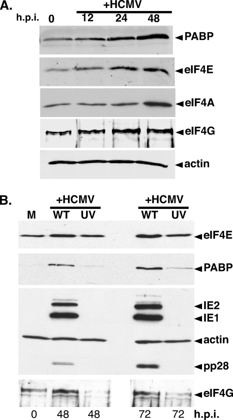
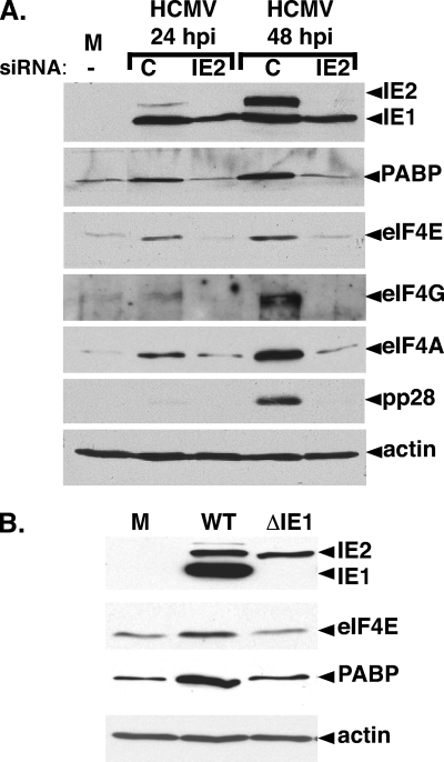
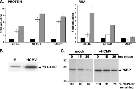
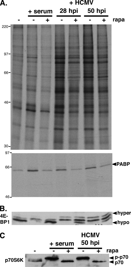
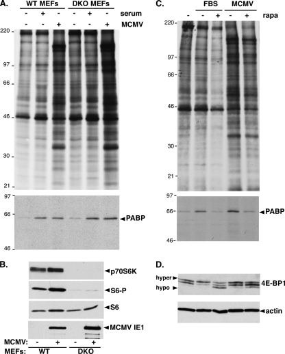
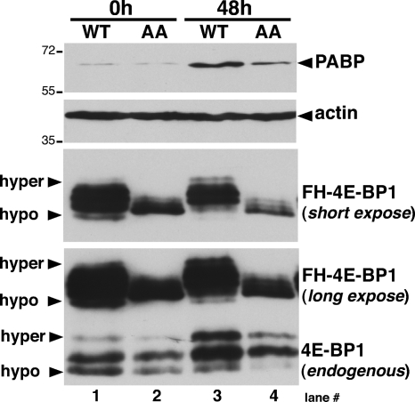
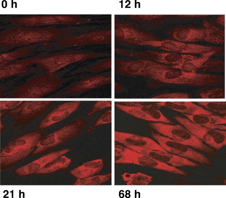
Irrespective of their effects on ongoing host protein synthesis, productive replication of the representative alphaherpesvirus herpes simplex virus type 1, the representative gammaherpesvirus Kaposi's sarcoma herpesvirus, and the representative betaherpesvirus human cytomegalovirus [HCMV] stimulates the assembly of the multisubunit, cap-binding translation factor eIF4F. However, only HCMV replication is associated with an increased abundance of eIF4F core components (eIF4E, eIF4G, eIF4A) and the eIF4F-associated factor poly(A) binding protein (PABP). Here, we demonstrate that the increase in translation factor concentration was readily detected in an asynchronous population of HCMV-infected primary human fibroblasts, abolished by prior UV inactivation of virus, and genetically dependent upon viral immediate-early genes. Strikingly, while increased mRNA steady-state levels accompanied the rise in eIF4E and eIF4G protein levels, the overall abundance of PABP mRNA, together with the half-life of the polypeptide it encodes, remained relatively unchanged by HCMV infection. Instead, HCMV-induced PABP accumulation resulted from new protein synthesis and was sensitive to the mTORC1-selective inhibitor rapamycin, which interferes with phosphorylation of the mTORC1 substrate p70 S6K and the translational repressor 4E-BP1. While virus-induced PABP accumulation did not require p70 S6K, it was inhibited by the expression of a dominant-acting 4E-BP1 variant unable to be inactivated by mTORC1. Finally, unlike the situation in alpha- or gammaherpesvirus-infected cells, where PABP is redistributed to nuclei, PABP accumulated in the cytoplasm of HCMV-infected cells. Thus, cytoplasmic PABP accumulation is translationally controlled in HCMV-infected cells via a mechanism requiring mTORC1-mediated inhibition of the cellular 4E-BP1 translational repressor.


Figures

FIG. 1.
FIG. 1.
Elevation of the translation initiation factor concentration in HCMV-infected cells requires viral gene expression. (A) Asynchronous, subconfluent NHDFs were mock infected (0 h.p.i.) or infected with HCMV. At the indicated times postinfection, total protein was isolated, fractionated by SDS-PAGE, and analyzed by immunoblotting with the indicated antisera. (B) UV inactivation of HCMV abrogates the increase in translation initiation factor levels. NHDFs were either mock infected (M) or infected (MOI = 5) with WT or UV-inactivated (UV) HCMV. At the indicated times postinfection, total protein was analyzed by immunoblotting as described for panel A.
FIG. 2.
FIG. 2.
HCMV-mediated translation factor accumulation is dependent upon viral IE gene expression. (A) Inhibition of translation factor accumulation by RNAi-mediated IE2 silencing. NHDFs transiently transfected with control noninterfering siRNA (C), IE2 siRNA (IE2), or no siRNA (−) were either mock infected (M) or infected with HCMV (MOI = 5). Total protein was harvested at the indicated times postinfection and analyzed by immunoblotting with the indicated antisera. (B) Quiescent NHDFs were either mock infected (M) or infected with WT HCMV (Towne strain) or an IE1-deficient mutant (ΔIE1). Total protein was harvested at 60 hpi and analyzed by immunoblotting as described for panel A.
FIG. 3.
FIG. 3.
Translational control of PABP levels in HCMV-infected cells. (A) Analysis of eIF4E, eIF4G, and PABP mRNA abundance in HCMV-infected cells. Serum-starved NHDFs were mock infected (0 h) or infected with HCMV (MOI = 5). At 0 hpi (black bars), 70 hpi (white bars), and 96 hpi (gray bars), total protein (left) was fractionated by SDS-PAGE and analyzed by immunoblotting with the indicated antisera. Chemiluminescence images were captured and quantified with a Bio-Rad ChemiDoc XR5 system. Total RNA (right) was isolated, and eIF4G1, eIF4E, and PABP mRNA levels were determined by real-time RT-PCR (normalized to actin). (B) Stimulation of new PABP synthesis in HCMV-infected cells. NHDFs were infected as described for panel A. At 24 hpi, cultures were pulse-labeled for 2 h with [35S]Met-Cys, and PABP was immunoprecipitated. Immune complexes were fractionated by SDS-PAGE and visualized by autoradiography. (C) PABP stability is similar in both mock- and HCMV-infected cells. NHDFs infected as described for panel A were pulse-labeled at 50 hpi as described for panel B and then returned to unlabeled medium for the indicated chase times. Total protein was subsequently isolated, and PABP was immunoprecipitated and analyzed as described for panel B. The mobility of molecular mass standards (in kilodaltons) is shown to the left. The percentage of [35S]PABP remaining over time was quantified by densitometry. In each case (mock versus HCMV infection), the amount of radiolabeled PABP after the pulse (0 h chase) was set at 100%.
FIG. 4.
FIG. 4.
Rapamycin sensitivity of HCMV-induced PABP mRNA translation and p70RSK activation. (A, top) Growth-arrested NHDFs were either serum stimulated (+ serum) or infected with HCMV (MOI = 5). At 26 or 46 hpi, HCMV-infected cells were treated with either DMSO or rapamycin (rapa) for 30 min and subsequently pulse-labeled with [35S]Met-Cys with (+) or without (−) rapa for 1.5 h. Uninfected cells (with or without rapa) were similarly metabolically labeled following stimulation with 20% FBS for 20 min. Total protein (top) and PABP immunoprecipitates (bottom) were fractionated by SDS-PAGE and visualized by autoradiography. The migration of molecular mass standards (in kilodaltons) is shown on the left. (B, C) Lysates from panel A were fractionated by SDS-PAGE and analyzed by immunoblotting using the indicated antibodies. The hyper- and hypophosphorylated 4E-BP1 forms are designated. p-p70, phosphorylated p70 S6K.
FIG. 5.
FIG. 5.
Induction of PABP mRNA translation by MCMV is preserved in p70 S6K-deficient cells. (A) Growth-arrested WT or p70 S6K1/K2 doubly deficient primary MEFs (DKO) were mock infected (−), infected (+) with MCMV (MOI = 5), or serum stimulated. At 24 hpi, cultures were metabolically labeled with [35S]Met-Cys for 2 h. Cell-free lysates (top) and PABP immunoprecipitates (bottom) were fractionated by SDS-PAGE and analyzed by autoradiography. The mobility of molecular weight standards (in kilodaltons) is shown on the left. (B) Total protein isolated from MEFs infected as described for panel A was fractionated by SDS-PAGE and analyzed by immunoblotting with the indicated antibodies. S6-P, phospho-specific S6. (C) At 22 hpi, MEFs (infected as described for panel A) were pulse-labeled with [35S]Met-Cys for 2 h with or without rapamycin. Cell-free lysates (top) and PABP immunoprecipitates (bottom) were analyzed as described for panel A. FBS, fetal bovine serum. (D) Samples described in panel C were analyzed by immunoblotting with the indicated antisera. The hyper- and hypophosphorylated forms of 4E-BP1 are indicated.
FIG. 6.
FIG. 6.
Regulation of PABP accumulation by 4E-BP1 phosphorylation in HCMV-infected cells. NHDFs stably expressing epitope-tagged (FH) WT or AA mutant 4E-BP1 were growth arrested by serum deprivation and either mock infected (0 h) or infected with HCMV (MOI = 5). After 48 h, total protein was isolated, fractionated by SDS-PAGE, and analyzed by immunoblotting with anti-4E-BP1 antibody which recognizes both endogenous and ectopically expressed (FH-4E-BP1) forms. Long and short exposures of the ectopically expressed 4E-BP1 protein are shown.
FIG. 7.
FIG. 7.
Cytoplasmic accumulation of PABP in HCMV-infected cells. NHDFs were either mock infected (0 h) or infected with HCMV (MOI = 5). At the indicated times postinfection (12 h, 21 h, 68 h), cells were fixed and processed for indirect immunofluorescence assay using anti-PABP.

References

    1. Afonina, E., R. Stauber, and G. N. Pavlakis. 1998. The human poly(A)-binding protein 1 shuttles between the nucleus and the cytoplasm. J. Biol. Chem. 273:13015-13021. - PubMed
    1. Arias, C., D. Walsh, J. Harbell, A. C. Wilson, and I. Mohr. 2009. Activation of host translational control pathways by a viral developmental switch. PLoS Pathog. 5:e1000334. - PMC - PubMed
    1. Braunstein, S., K. Karpisheva, C. Pola, J. Goldberg, T. Hochman, H. Yee, J. Cangiarella, R. Arju, S. C. Formenti, and R. J. Schneider. 2007. A hypoxia-controlled cap-dependent to cap-independent translation switch in breast cancer. Mol. Cell 28:501-512. - PubMed
    1. Buchkovich, N. J., Y. Yu, C. A. Zampieri, and J. C. Alwine. 2008. The TORrid affairs of viruses: effects of mammalian DNA viruses on the PI3K-Akt-mTOR signaling pathway. Nat. Rev. Microbiol. 6:266-275. - PMC - PubMed
    1. Dobrikova, E., M. Shveygert, R. Walters, and M. Gromeier. 2010. Herpes simplex virus proteins ICP27 and UL47 associate with polyadenylate-binding protein and control its sub-cellular distribution. J. Virol. 84:270-279. - PMC - PubMed

Publication types

MeSH terms

LinkOut - more resources