. 2011 Jan;85(1):644-8.
doi: 10.1128/JVI.01851-10. Epub 2010 Oct 27.

R5 HIV env and vesicular stomatitis virus G protein cooperate to mediate fusion to naive CD4+ T Cells

Affiliations

R5 HIV env and vesicular stomatitis virus G protein cooperate to mediate fusion to naive CD4+ T Cells

Matthew J Pace et al. J Virol. 2011 Jan.

Abstract

Naïve CD4(4) T cells are resistant to both HIV R5 env and vesicular stomatitis virus G protein (VSV-G)-mediated fusion. However, viral particles carrying both HIV R5 env and VSV-G infect naïve cells by an unexplained mechanism. We show that VSV-G-pseudotyped virus cannot fuse to unstimulated cells because the viral particles cannot be endocytosed. However, virions carrying both HIV R5 env and VSV-G can fuse because CD4 binding allows viral uptake. Our findings reveal a unique mechanism by which R5 HIV env and VSV-G cooperate to allow entry to naïve CD4(+) T cells, providing a tool to target naïve CD4(+) T cells with R5 HIV to study HIV coreceptor signaling and latency.


Figures

FIG. 1.
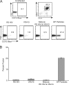
FIG. 1.
DP particles with R5 HIV env and VSV-G overcome a fusion restriction in naïve CD4+ T cells. A total of 5 × 10⁵ unstimulated CD4+ T cells were spinoculated with 8 μg/ml Polybrene with viruses containing BlamVpr. R5 HIV was generated by transfecting pNLAD8 (13), and VSV-G-pseudotyped particles were generated by transfecting pNL4-3Δenv and pHIT as previously described (2). Double-pseudotyped (DP) particles were generated by transfecting pNLAD8 and pHIT. After spinoculation, cells were incubated for 1 h at 37°C to allow fusion. Cells were stained with anti-CD45RO Texas Red (Beckman Coulter) and anti-CCR7 PECy7 (BD Bioscience) to distinguish memory from naïve T cells. An uninfected control was used to set the gates in the infected samples. The numbers represent the percentages of cells in which fusion has occurred. All flow plots use a logarithmic scale. Representative fusion levels in one experiment are seen in panel A. The fusion levels of three separate experiments using three different donors are shown in panel B. The error bars represent the standard deviations of the measurements. The amount of virus added was based on late reverse transcripts found in CD3/CD28-activated CD4+ T cells after 24 h. FSC, forward scatter.
FIG. 2.
FIG. 2.
DP particles can fuse in a coreceptor-independent manner. A total of 5 × 10⁵ CD4+ T cells from a Δ32 donor, whose cells do not express CCR5, were infected, and a fusion assay was performed as described for Fig. 1. CD4+ T cells were labeled with anti-CD3 allophycocyanin (APC) and anti-CD4 PerCpCy5.5 (BD Bioscience) to identify CD4+ T cells. Data were analyzed as described for Fig. 1. Representative fusion levels in one experiment are shown in panel A. The fusion levels of three separate experiments are shown in panel B. The error bars represent the standard deviations of the measurements. FSC, forward scatter.
FIG. 3.
FIG. 3.
DP particles require CD4 binding and low pH but not HIV env-mediated fusion to fuse to naïve CD4+ T cells. A total of 5 × 10⁵ unstimulated CD4+ T cells were left untreated, preincubated with 10 mM NH4Cl for 1 h at room temperature, preincubated with 25 μg/ml anti-CD4 clone 19 (Ron Collman) for 1 h at 4°C, or treated with 10 μg/ml T20 (AIDS Research and Reference Reagent Program) immediately before spinoculation. All cells were then spinoculated with 8 μg/ml Polybrene by using NL4-3, an X4-tropic HIV strain, or DP particles, and a BlamVpr fusion assay was performed as described for Fig. 1. X4 HIV was generated by transfecting pNL4-3 as previously described (2). Only fusion to naïve cells is shown. Numbers represent the mean fusion percentages relative to the values for untreated controls for three separate experiments using three different donors. Error bars represent the standard deviations of the measurements.
FIG. 4.
FIG. 4.
There is a VSV-G-mediated endocytosis restriction in unstimulated CD4+ T cells. A total of 4 × 10⁶ unstimulated CD4+ T cells were spinoculated with R5 HIV, DP particles, VSV-G-pseudotyped particles (VSV-G), and particles with no envelope (generated by transfecting with pNL4-3Δenv) at 4°C. After unbound virions were washed away, samples were collected as follows. Half of the remaining cells were placed on ice and treated with 10 mg/ml pronase (Roche) to remove noninternalized virions (referred to as the cold sample). The other half of the cells were incubated at 37°C for 2 h to allow viral uptake (referred to as the warm sample). After the warm sample was incubated for 2 h, the cells were treated with pronase. Virions still associated with the cells after pronase treatments in both cold and warm samples were measured via a p24 enzyme-linked immunosorbent assay (ELISA). The amount of internalized virions was then calculated by subtracting the amount of p24 associated with the cold fraction from the amount of p24 associated with the warm fraction. The amount of p24 was then converted to virions/cell as previously described (27). The graph reflects three different inoculations with different donors. The error bars represent the standard deviations of the measurements.

References

    1. Agosto, L. M., J. J. Yu, J. Dai, R. Kaletsky, D. Monie, and U. O'Doherty. 2007. HIV-1 integrates into resting CD4+ T cells even at low inoculums as demonstrated with an improved assay for HIV-1 integration. Virology 368:60-72. - PMC - PubMed
    1. Agosto, L. M., J. J. Yu, M. K. Liszewski, C. Baytop, N. Korokhov, L. M. Humeau, and U. O'Doherty. 2009. The CXCR4-tropic human immunodeficiency virus envelope promotes more-efficient gene delivery to resting CD4+ T cells than the vesicular stomatitis virus glycoprotein G envelope. J. Virol. 83:8153-8162. - PMC - PubMed
    1. Amara, A., A. Vidy, G. Boulla, K. Mollier, J. Garcia-Perez, J. Alcami, C. Blanpain, M. Parmentier, J. L. Virelizier, P. Charneau, and F. Arenzana-Seisdedos. 2003. G protein-dependent CCR5 signaling is not required for efficient infection of primary T lymphocytes and macrophages by R5 human immunodeficiency virus type 1 isolates. J. Virol. 77:2550-2558. - PMC - PubMed
    1. Atchison, R. E., J. Gosling, F. S. Monteclaro, C. Franci, L. Digilio, I. F. Charo, and M. A. Goldsmith. 1996. Multiple extracellular elements of CCR5 and HIV-1 entry: dissociation from response to chemokines. Science 274:1924-1926. - PubMed
    1. Bosque, A., and V. Planelles. 2009. Induction of HIV-1 latency and reactivation in primary memory CD4+ T cells. Blood 113:58-65. - PMC - PubMed

Publication types

MeSH terms

Substances