. 2010 Oct 27;30(43):14476-81.
doi: 10.1523/JNEUROSCI.1698-10.2010.

Oligodendrocyte myelin glycoprotein does not influence node of ranvier structure or assembly

Affiliations

Oligodendrocyte myelin glycoprotein does not influence node of ranvier structure or assembly

Kae-Jiun Chang et al. J Neurosci. .

Abstract

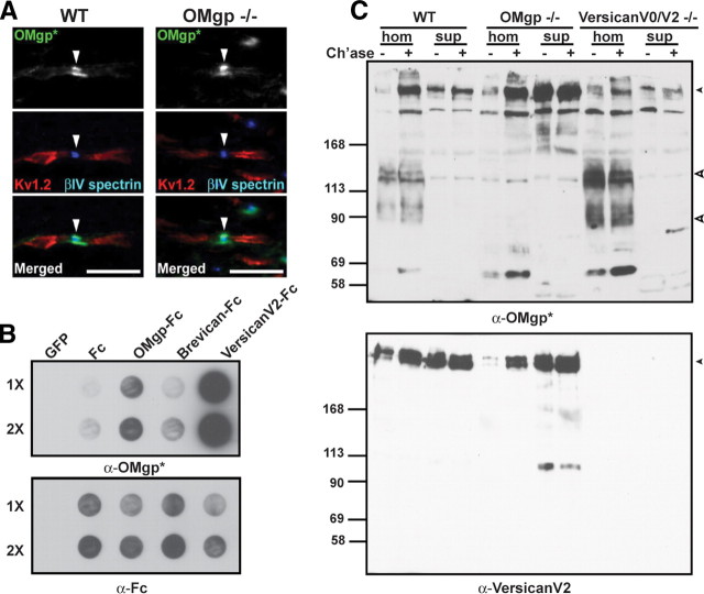
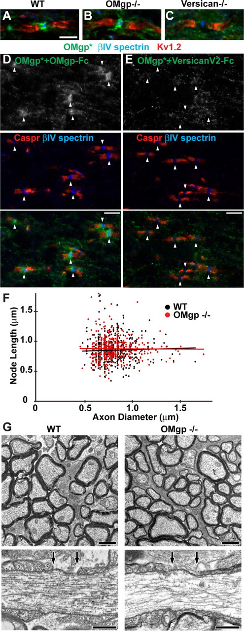
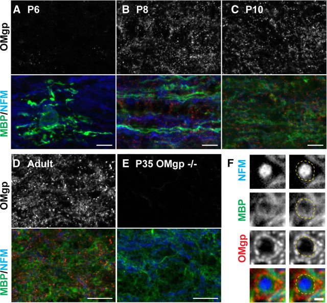
Oligodendrocyte myelin glycoprotein (OMgp) is expressed by both neurons and oligodendrocytes in the CNS. It has been implicated in growth cone collapse and neurite outgrowth inhibition by signaling through the Nogo receptor and paired Ig-like receptor B (PirB). OMgp was also reported to be an extracellular matrix (ECM) protein surrounding CNS nodes of Ranvier and proposed to function as (1) an inhibitor of nodal collateral sprouting and (2) an important contributor to proper nodal and paranodal architecture. However, we show here that the anti-OMgp antiserum used in previous studies to define the functions of OMgp at nodes is not specific. Among all reported nodal ECM components, the antiserum exhibited strong cross-reactivity against versican V2 isoform, a chondroitin sulfate proteoglycan. Furthermore, the OMgp antiserum labeled OMgp-null nodes, but not nodes from versican V2-deficient mice, and preadsorption of the OMgp antiserum with recombinant versican V2 blocked nodal labeling. Analysis of CNS nodes in OMgp-null mice failed to reveal any nodal or paranodal defects, or increased nodal collateral sprouting, indicating that OMgp does not participate in CNS node of Ranvier assembly or maintenance. We successfully identified a highly specific anti-OMgp antibody and observed OMgp staining in white matter only after initiation of myelination. OMgp immunoreactivity decorated the surface of mature myelinated axons, but was excluded from compact myelin and nodes. Together, our results strongly argue against the nodal localization of OMgp and its proposed functions at nodes, and reveal OMgp's authentic localization relative to nodes and myelin.


Figures

Figure 1.
Figure 1.
α-OMgp* is not specific to OMgp and cross-reacts with versican V2. A, Spinal cord sections from wild-type and OMgp-null mice immunostained with α-OMgp* (green), anti-βIV spectrin (blue), and anti-Kv1.2 (red). Arrowheads indicate nodes of Ranvier. Scale bars, 10 μm. B, Media collected from COS-7 cell cultures expressing palmitoylated EGFP (GFP), Fc, OMgp-Fc, brevican-Fc, and versican V2-Fc were applied to nitrocellulose membranes onefold (1×) or twofold (2×) concentrated and probed with α-OMgp* or anti-human Fc. C, Immunoblotting of brain membrane homogenates (hom; 75 μg) and cytosolic fractions (sup; 20 μg) made from WT, OMgp-null, and versican V0/V2-null brains with or without chondroitinase ABC treatment (Ch'ase +/−). Blots were probed with α-OMgp* or anti-versican V2 antibodies. The top arrowhead indicates the position of versican V2, and the bottom two open arrowheads indicate OMgp with (upper) and without (lower) the GPI moiety (Mikol and Stefansson, 1988).
Figure 2.
Figure 2.
α-OMgp* detected versican V2 at CNS nodes of Ranvier. A–C, Optic nerve sections of WT, OMgp-null, and versican V0/V2-null mice stained with α-OMgp* (green), anti-βIV spectrin (blue), and anti-Kv1.2 (red). Scale bar, 5 μm. D, E, α-OMgp* was preadsorbed with OMgp-Fc (D) or versican V2-Fc (E) before being incubated with WT spinal cord sections. Arrowheads indicate nodes of Ranvier. Scale bars, 5 μm. F, Node length plotted as a function of axon diameter in WT and OMgp-null optic nerves. G, Electron micrographs of optic nerves from 100-d-old wild-type and OMgp null (−/−) mice. Top panels show cross sections of myelinated axons in the optic nerves. Lower panels show single nodes of Ranvier from longitudinal sections of optic nerves. The nodal gap is defined by the two arrows above each axon. Scale bars: top, 1 μm; bottom, 0.5 μm.
Figure 3.
Figure 3.
A specific anti-OMgp antibody detected no accumulation of OMgp at CNS nodes of Ranvier. A, Media collected from COS-7 cell cultures expressing palmitoylated EGFP (GFP), Fc, OMgp-Fc, brevican-Fc, and versican V2-Fc were applied to nitrocellulose membranes onefold (1×) or twofold (2×) concentrated and probed with a goat anti-OMgp antibody or anti-human Fc antibody. B, Immunoblotting of brain membrane homogenates (hom; 75 μg) and cytosolic fractions (sup; 20 μg) made from WT, OMgp-null, and versican V0/V2-null brains with or without chondroitinase ABC treatment (Ch'ase +/−). Blots were probed with goat anti-OMgp, stripped, and reprobed using anti-versican V2 antibodies. C, Western blotting of wild-type (+) and OMgp-null (−) brain membrane homogenates (Br; 10 μg) and optic nerve (ON; 10 μg) and sciatic nerve (20 μg) extracts with anti-MBP and goat anti-OMgp. D, WT and OMgp-null spinal cord cross sections stained with goat anti-OMgp. E, WT and OMgp-null spinal cord longitudinal sections immunostained with goat anti-OMgp (green), anti-Caspr (red), and anti-βIV spectrin (blue). Arrowheads indicate nodes. Scale bars, 10 μm.
Figure 4.
Figure 4.
OMgp is expressed during myelination and localizes outside compact myelin. A–E, Immunostaining of wild-type P6 (A), P8 (B), P10 (C), and adult (D) and OMgp-null P35 (E) optic nerves using goat anti-OMgp (red), anti-MBP (green), and anti-neurofilament M (NFM). Scale bars, 10 μm. F, Immunostaining of adult wild-type spinal cord cross sections using goat anti-OMgp (red), anti-MBP (green), and anti-NFM (blue). The dotted line indicates the outer surface of the compact myelin. Scale bar, 1 μm.

References

    1. Apostolski S, Sadiq SA, Hays A, Corbo M, Suturkova-Milosevic L, Chaliff P, Stefansson K, LeBaron RG, Ruoslahti E, Hays AP, Latov N. Identification of Gal(β1–3)GalNAc bearing glycoproteins at the nodes of Ranvier in peripheral nerve. J Neurosci Res. 1994;38:134–141. - PubMed
    1. Atwal JK, Pinkston-Gosse J, Syken J, Stawicki S, Wu Y, Shatz C, Tessier-Lavigne M. PirB is a functional receptor for myelin inhibitors of axonal regeneration. Science. 2008;322:967–970. - PubMed
    1. Bartsch U, Pesheva P, Raff M, Schachner M. Expression of janusin (J1–160/180) in the retina and optic nerve of the developing and adult mouse. Glia. 1993;9:57–69. - PubMed
    1. Bekku Y, Vargová L, Goto Y, Vorísek I, Dmytrenko L, Narasaki M, Ohtsuka A, Fässler R, Ninomiya Y, Syková E, Oohashi T. Bral1: its role in diffusion barrier formation and conduction velocity in the CNS. J Neurosci. 2010;30:3113–3123. - PMC - PubMed
    1. Berghs S, Aggujaro D, Dirkx R, Jr, Maksimova E, Stabach P, Hermel JM, Zhang JP, Philbrick W, Slepnev V, Ort T, Solimena M. betaIV spectrin, a new spectrin localized at axon initial segments and nodes of Ranvier in the central and peripheral nervous system. J Cell Biol. 2000;151:985–1002. - PMC - PubMed

Publication types

MeSH terms

LinkOut - more resources