. 2010 Oct 27;30(43):14543-51.
doi: 10.1523/JNEUROSCI.3037-10.2010.

Locus ceruleus and anterior cingulate cortex sustain wakefulness in a novel environment

Affiliations

Locus ceruleus and anterior cingulate cortex sustain wakefulness in a novel environment

Heinrich S Gompf et al. J Neurosci. .

Abstract

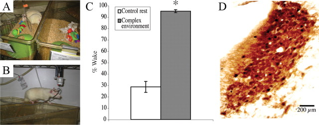
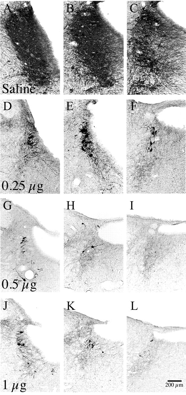
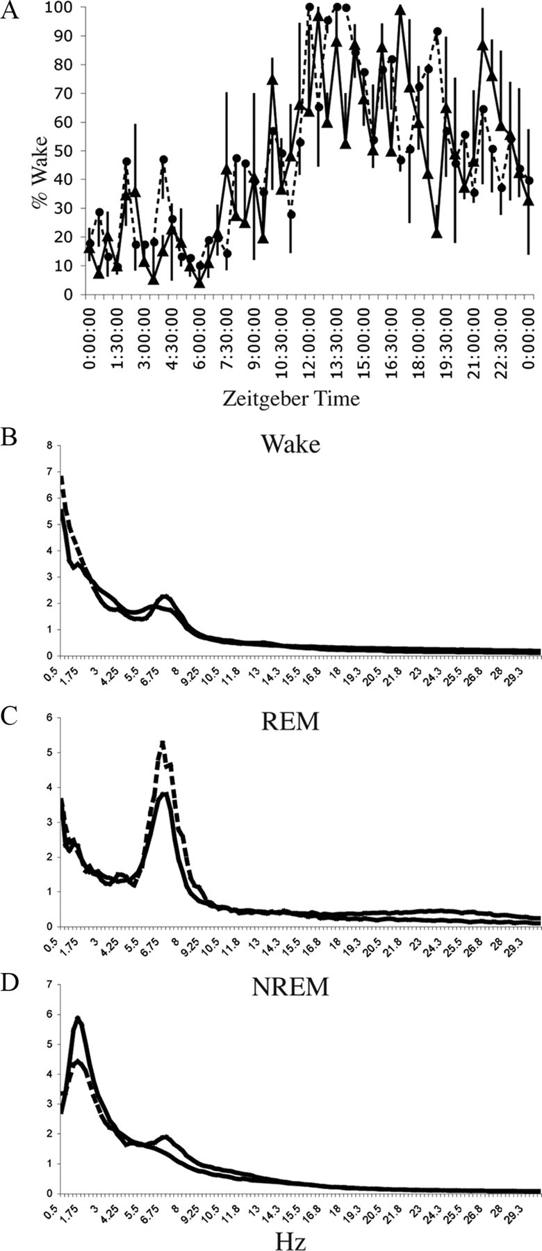
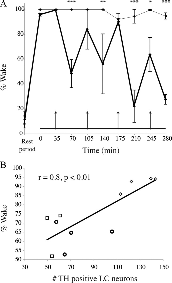
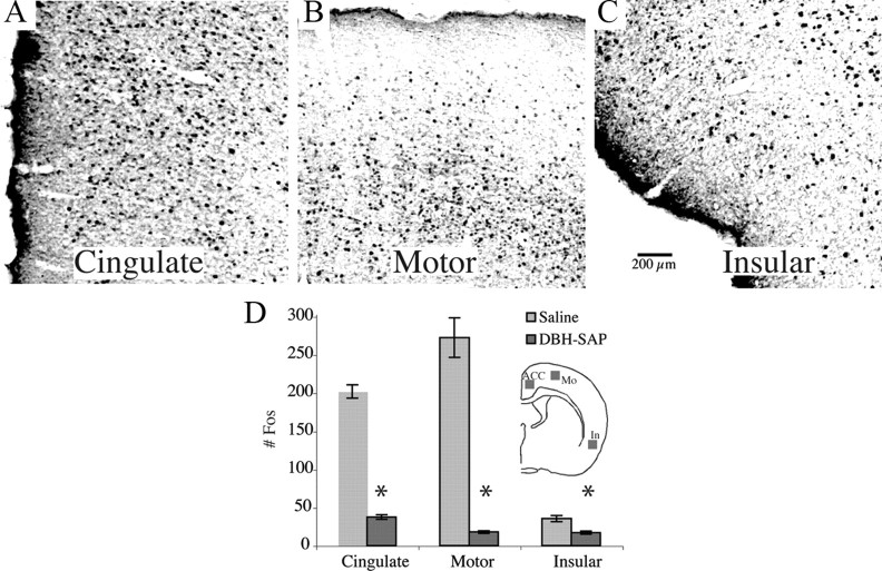
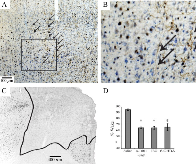
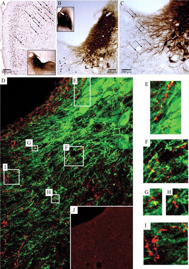
Locus ceruleus (LC) neuronal activity is correlated with the waking state, yet LC lesions produce only minor alterations in daily wakefulness. Here, we report that sustained elevations in neurobehavioral and EEG arousal in response to exposure to an environment with novel stimuli, including social interaction, are prevented by selective chemical lesions of the LC in rats. Similar results are seen when the anterior cingulate cortex (ACC), which receives especially dense LC innervation, is selectively denervated of LC input or is ablated by the cell-specific neurotoxin ibotenic acid. Anterograde tracing combined with tyrosine hydroxylase immunohistochemistry demonstrates ACC terminals in apposition with the distal dendrites of LC neurons. Our data implicate the ACC as both a source of input to the LC as well as one of its targets and suggests that the two structures engage in a dialog that may provide a critical neurobiological substrate for sustained attention.


Figures

Figure 1.
Figure 1.
Wakefulness in a complex environment. A, B, Example of complex environment. Two rats were paired by bringing together two regular housing cages with tops removed (A) so that they were free to move between cages (B) and interact. Novel objects were added—baby rattles and plastic cones are shown in A—every 70 min for 5 h to arouse curiosity and exploration. C, Animals spent significantly (*p = 1 × 10⁻⁷) more time awake in this environment than during the same period 24 h prior in their home cages. D, Double immunohistochemistry for tyrosine hydroxylase (brown cytoplasm) and c-Fos (black nuclei) at the level of the LC. LC c-Fos expression, usually absent during baseline wakefulness, increased dramatically during novelty exposure.
Figure 2.
Figure 2.
AL, A series of photomicrographs showing loss of TH-immunoreactive neurons in the LC at three levels, after injection of saline (A–C) or increasing doses (DF, 0.25 μg; GI, 0.5 μg; JL, 1 μg) of α-DBH-SAP into the lateral ventricle. Note the substantial reduction in TH-positive LC neurons in all α-DBH-SAP-treated groups relative to control saline injections, and that the cell loss following 0.5 and 1.0 μg injections was near maximal.
Figure 3.
Figure 3.
LC lesions do not affect baseline wakefulness. A, Percentage wake time ± SEM per 30 min period in their home cage on the baseline recording day for intact animals (n = 3, solid line) and animals with LC lesions due to injection of 0.5 μg of α-DBH-SAP into the lateral ventricle (n = 3, dashed line). Time is shown with respect to lights on (7 A.M., Zeitgeber time). No significant differences were found (p = 0.9574, F = 0.2115, n = 3 each). B–D, Power spectra for control (n = 3, solid line) and 0.5 μg of α-DBH-SAP-treated (n = 3, dashed line) animals for the wake (B), REM sleep (C), and slow-wave sleep (D) stages over frequency ranges from 0.5 to 30 Hz. No significant differences were found at any frequency in any stage (repeated-measures ANOVA, p > 0.9 for each point).
Figure 4.
Figure 4.
Reduced wakefulness during novelty exposure in LC-lesioned animals compared with saline-injected controls. A, Averaged total wakefulness of 0.5 μg of α-DBH-SAP-treated animals (n = 4, thick line) compared with saline-injected controls (thin line) in 35 min bins over the 5 h novelty exposure. The first data point represents wakefulness in the 35 min period just before the complex environment period (horizontal line at bottom). Arrows indicate times of presentation of new sets of novel objects. Note that saline-treated control animals maintain almost full wakefulness, whereas α-DBH-SAP-treated animals show increased wakefulness bouts around the time of novel object presentation, which is then markedly decreased in the second half of each 70 min period. This trend was more pronounced the longer animals were exposed to environmental complexity. Points are means ± SEM. *p < 0.05; **p < 0.01; ***p < 0.001. B, Correlation between the number of LC neurons and wakefulness in the complex environment. Diamonds, 0.25 μg of α-DBH-SAP; circles, 0.5 μg of α-DBH-SAP; squares, 1 μg of α-DBH-SAP.
Figure 5.
Figure 5.
Cortical activation in a complex environment. AC, c-Fos expression was increased in all cortical areas after 5 h of sustained wakefulness during novelty exposure, e.g., the anterior cingulate (A), motor (B), and insular cortices (C). D, Elevated cortical c-Fos was maintained throughout the 5 h period in saline-injected animals. However, α-DBH-SAP-treated animals exhibited marked decreases in cortical c-Fos expression. *p < 0.001. Inset, Approximate location of 1 × 1 mm counting boxes. Mo, Motor cortex; In, insular cortex.
Figure 6.
Figure 6.
Cortical c-Fos activation is not correlated with noradrenergic fiber loss. A, B, 6-OHDA unilaterally injected into the LC produced widespread LC neuronal loss (A), accompanied by widespread loss of DBH-immunoreactive fibers in the ipsilateral cerebral cortex (B). C, Five hours of sustained wakefulness during novelty exposure evoked widespread c-Fos expression in the ACC, both ipsilateral and contralateral to the LC lesion.
Figure 7.
Figure 7.
Anterior cingulate cortex involvement in wakefulness induced by a complex environment. A, A photomicrograph showing the border of an injection of 6-OHDA into the ACC (left half) and relatively normal dopamine-β-hydroxylase-immunoreactive innervation of the adjacent premotor cortex (fine brown axons on right half, indicated by arrows). The sample was Nissl stained (blue cell bodies) and immunostained for c-Fos (black nuclei). B, Boxed area in A, magnified to show the transition from lesioned to unlesioned in more detail. C, ACC lesion (boundaries illustrated by black line) resulting from ibotenate injection into same area as A. D, For comparison, total wakefulness over the 5 h period of exposure to the complex environment for animals treated with saline or α-DBH-SAP intraventricularly, or with ibotenic acid (IBO) or 6-OHDA injections into the ACC. No significant differences (*p = 0.9) were found between α-DBH-SAP, ibotenic acid, and 6-OHDA-injected animals, indicating that the LC expresses its effects on wakefulness via its inputs to the ACC.
Figure 8.
Figure 8.
Anterior cingulate cortex provides afferents to the LC. A, CTb injected into the LC and the area just medial to it (inset) retrogradely labeled neurons in the ACC (arrows distinguish brown retrogradely labeled neurons from black nuclear Fos staining in the same experiment). B, C, Low- and high-magnification images of the LC after injection of biotinylated dextranamine into the ACC (B, inset) showed evidence of numerous black axon terminals (B, arrowheads) adjacent to brown TH-immunopositive dendrites projecting medially from the LC (C, arrows). D, This relationship is best seen with confocal microscopy in an immunofluorescence image from the same case, showing biotinylated dextranamine-stained anterior cingulate axon terminals (red) and tyrosine hydroxylase-positive LC dendrites (green). In this 15-μm-thick z-stack composed of twenty 0.75-μm-thick individual images, boxes correspond to the locations of the enlarged images in EI. Note the yellow areas where overlap between green and red indicates appositions. Appositions were confirmed by examining individual single plane images. A magenta–green copy of EJ is available as supplemental Figure 1, available at www.jneurosci.org as supplemental material. J, The inset shows the same field as D, but with only the red channel to indicate the substantial ACC innervation of this region.

References

    1. Aston-Jones G, Bloom FE. Activity of norepinephrine-containing locus coeruleus neurons in behaving rats anticipates fluctuations in the sleep-waking cycle. J Neurosci. 1981;1:876–886. - PMC - PubMed
    1. Aston-Jones G, Cohen JD. An integrative theory of locus coeruleus-norepinephrine function: adaptive gain and optimal performance. Annu Rev Neurosci. 2005;28:403–450. - PubMed
    1. Aston-Jones G, Ennis M, Pieribone VA, Nickell WT, Shipley MT. The brain nucleus locus coeruleus: restricted afferent control of a broad efferent network. Science. 1986;234:734–737. - PubMed
    1. Berridge CW, España RA. Synergistic sedative effects of noradrenergic alpha(1)- and beta-receptor blockade on forebrain electroencephalographic and behavioral indices. Neuroscience. 2000;99:495–505. - PubMed
    1. Berridge CW, Foote SL. Effects of locus coeruleus activation on electroencephalographic activity in neocortex and hippocampus. J Neurosci. 1991;11:3135–3145. - PMC - PubMed

Publication types

MeSH terms

Substances