. 2010 Nov 16;107(46):19921-6.
doi: 10.1073/pnas.1007256107. Epub 2010 Oct 27.

Inositol hexakisphosphate kinase-2 acts as an effector of the vertebrate Hedgehog pathway

Affiliations

Inositol hexakisphosphate kinase-2 acts as an effector of the vertebrate Hedgehog pathway

Bhaskarjyoti Sarmah et al. Proc Natl Acad Sci U S A. .

Abstract

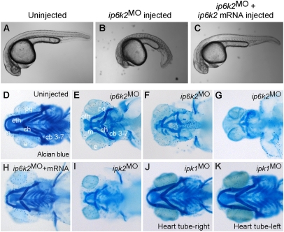
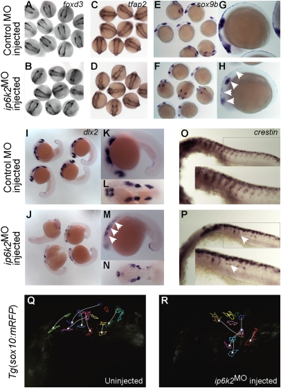
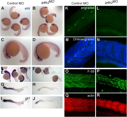
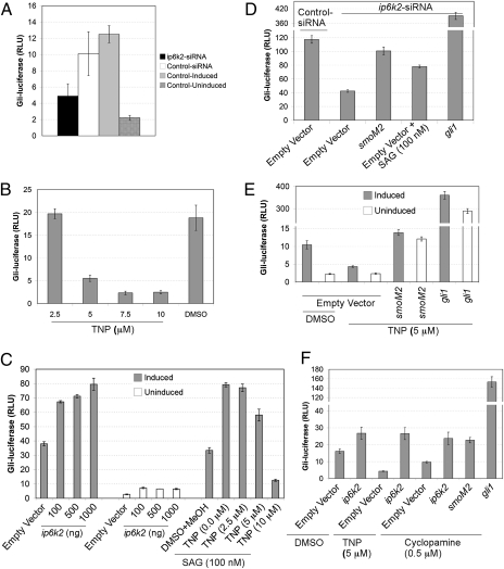
Inositol phosphate (IP) kinases constitute an emerging class of cellular kinases linked to multiple cellular activities. Here, we report a previously uncharacterized cellular function in Hedgehog (Hh) signaling for the IP kinase designated inositol hexakisphosphate kinase-2 (IP6K2) that produces diphosphoryl inositol phosphates (PP-IPs). In zebrafish embryos, IP6K2 activity was required for normal development of craniofacial structures, somites, and neural crest cells. ip6k2 depletion in both zebrafish and mammalian cells also inhibited Hh target gene expression. Inhibiting IP(6) kinase activity using N(2)-(m-(trifluoromethy)lbenzyl) N(6)-(p-nitrobenzyl)purine (TNP) resulted in altered Hh signal transduction. In zebrafish, restoring IP6K2 levels with exogenous ip6k2 mRNA reversed the effects of IP6K2 depletion. Furthermore, overexpression of ip6k2 in mammalian cells enhanced the Hh pathway response, suggesting IP6K2 is a positive regulator of Hh signaling. Perturbations from IP6K2 depletion or TNP were reversed by overexpressing smoM2, gli1, or ip6k2. Moreover, the inhibitory effect of cyclopamine was reversed by overexpressing ip6k2. This identified roles for the inositol kinase pathway in early vertebrate development and tissue morphogenesis, and in Hh signaling. We propose that IP6K2 activity is required at the level or downstream of Smoothened but upstream of the transcription activator Gli1.


Conflict of interest statement

The authors declare no conflict of interest.

Figures

Fig. 1.
Fig. 1.
IP6K2 activity is required for normal development of craniofacial and somite structures. (A–C) Lateral view of uninjected (A), ip6k2SPMO-injected (B), and ip6k2SPMO + ip6k2 mRNA-coinjected (C) live embryos at 24 hpf. Eighty percent of the ip6k2SPMO embryos had a small head and reduced trunk; both the defects were rescued by ip6k2 mRNA coinjection. Effective ip6k2SPMO suppression of splicing was confirmed by RT-PCR (Fig. S3 A and B). (D–K) Ventral view of Alcian blue stained head skeleton of 5-d-old uninjected control (D), ip6k2SPMO (E–G), ip6k2SPMO + ip6k2 mRNA (H), ipk2SPMO (I), and ipk1MO1 (J and K)-injected embryos. The anterior neurocranium was mostly maintained in 40% of ip6k2MO embryos, but distorted or reduced in 60% of embryos (F and G). The pharyngeal skeleton was affected in 92% embryos injected (n = 90). ip6k2 mRNA coinjection restored normal craniofacial skeleton (H). Craniofacial skeleton was altered in ipk2SPMO embryos (I). No craniofacial deformity was evident in ipk1MO1 embryos (J and K). e, eye; m, Meckel's cartilage; pq, palatoquadrate; ch, ceratohyal; cb 3–7, ceratobranchial arches 3–7; eth, ethmoid plate.
Fig. 2.
Fig. 2.
IP6K2 is critical for NCC development and migration. (A–P) Whole-mount in situ hybridizations for NC marker gene expression specifying distinct stages of NC development: (A–D) specification (foxd3 and tfap2 at 11 hpf), (E and H) maintenance (sox9b at 14 hpf), and (I–P) migration (dlx2 and crestin, 22 hpf) in control MO (A, C, E, G, I, K, L, and O) and ip6k2ATGMO (B, D, F, H, J, M, N, and P)-injected embryos. Arrowheads indicate distinct differences in the expression of sox9b (E–H), dlx2 (I–N), and crestin (O and P) between control MO and ip6k2ATGMO embryos. (Q and R) Analysis of NCC migration in real time using Tg(sox10(7.2):mrfp) fish (17) (see SI Materials and Methods for details). Fluorescence images (frames) for the hindbrain and eye regions were captured from a 6-h time-lapse sequence, with dorsal toward the top and anterior to the left. The 0-min time point represents 16-hpf stage. Images of uninjected (Q) and ip6k2ATGMO-injected (R) embryos show an overlay of first and last images onto which the tracings of migratory paths of CNCC were superimposed. Arrowheads indicate end point of each cell's trajectory.
Fig. 3.
Fig. 3.
IP6K2 is required for Hh target gene expression during development. (A–J) Whole-mount in situ hybridizations showing expression of Hh target genes in zebrafish embryos (24-hpf stage) injected with either control MO (column 1) or ip6k2SPMO (column 2). Expression of shh (A–D) was slightly elevated, whereas ptc1 (E–H) and gli1 (I and J) expression was down-regulated in ip6k2SPMO (B, D, F, H, and J) compared with controls (A, C, E, G, and I). (K–N) Whole-mount immunohistochemistry showing engrailed protein levels (detected by 4D9 antibody) in control MO (K) or ip6k2SPMO (L) embryos at 24 hpf. Engrailed was not detected in ip6k2SPMO embryos (L). M and N overlay engrailed immunohistochemistry and TO-PRO3-stained DNA. (O–R) Lateral views of embryos at 24 hpf stained for myosin heavy chain using F59 antibody to visualize slow muscle cells (O and P) and F-actin using rhodamine-phalloidin to reveal both fast and slow muscle cells (Q and R). Dorsal is toward the top and anterior to the left.
Fig. 4.
Fig. 4.
IP6K2 positively regulates the Hh response pathway. (A) Depletion of ip6k2 reduces Shh-dependent Gli-luciferase activity. Bar graphs showing Gli-luciferase activity expressed as relative luciferase units (RLU) in NIH 3T3 cells cotransfected with 8× gli-luciferase and constitutive β-galactosidase reporter plasmids, and additionally subjected to ip6k2 or control siRNA treatment for 72 h. Cells were induced with Hh ligand (Shh-N conditioned medium) for 24 h, and Gli-luciferase activity was measured (normalized by β-galactosidase activity). Non–siRNA-treated cells (control induced) and non–siRNA-treated uninduced cells (control uninduced) were controls. (B) TNP inhibits Hh pathway activity. Shh-LIGHT Z3 cells were treated with TNP (2.5–10 μM) and Hh ligand induction for 24 h, and Gli-luciferase activity was quantified. DMSO treatment served as control. (C) IP6K2 acts as an effector of the Hh response pathway. Shh-LIGHT Z3 cells were transfected with an empty plasmid (1 μg) or a plasmid expressing ip6k2 ORF (0.1–1 μg) and assayed Gli-luciferase activity with and without Hh ligand induction after 24 h. Additionally, Shh-LIGHT Z3 cells were subjected to 100 nM SAG and increasing TNP concentrations (0.0–10 μM), with Gli-luciferase activity determined after 24 h induction with Hh ligand. (D) Epistasis analysis indicating IP6K2 acts at the level or downstream of Smo. Shh-LIGHT Z3 cells were cotransfected with control or ip6k2 siRNA and empty plasmid or plasmid expressing smoM2 or gli1, and 72 h later treated with Hh ligand alone or with SAG for 24 h. Gli-luciferase activity was determined. (E) smoM2 or gli1 overexpression rescues the inhibitory effect of TNP. NIH 3T3 cells were cotransfected with reporter plasmids, and either an empty plasmid or plasmid expressing smoM2 or gli1, grown for 24 h. Gli-luciferase activity was determined after 24-h treatment with TNP (5 μM) or DMSO, with or without Hh ligand induction. (F) ip6k2 overexpression reverses inhibitory effects of TNP and cyclopamine. NIH 3T3 cells were cotransfected with reporter plasmids and either an empty plasmid or plasmid expressing ip6k2, smoM2, or gli1, and grown for 24 h. Cells were subjected to Hh ligand induction in presence of DMSO, TNP (5 μM), or cyclopamine (0.5 μM), and Gli-luciferase activity was determined. Data are the mean ± SD for three independent assays.

References

    1. Varjosalo M, Taipale J. Hedgehog: Functions and mechanisms. Genes Dev. 2008;22:2454–2472. - PubMed
    1. Jiang J, Hui CC. Hedgehog signaling in development and cancer. Dev Cell. 2008;15:801–812. - PMC - PubMed
    1. Eaton S. Multiple roles for lipids in the Hedgehog signalling pathway. Nat Rev Mol Cell Biol. 2008;9:437–445. - PubMed
    1. Hausmann G, von Mering C, Basler K. The hedgehog signaling pathway: Where did it come from? PLoS Biol. 2009;7:e1000146. - PMC - PubMed
    1. Balla T, Szentpetery Z, Kim YJ. Phosphoinositide signaling: New tools and insights. Physiology (Bethesda) 2009;24:231–244. - PMC - PubMed

Publication types

MeSH terms

Substances