. 2010 Oct 19:1:97.
doi: 10.1038/ncomms1103.

A role for TREX components in the release of spliced mRNA from nuclear speckle domains

Affiliations

A role for TREX components in the release of spliced mRNA from nuclear speckle domains

Anusha P Dias et al. Nat Commun. .

Abstract

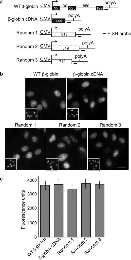
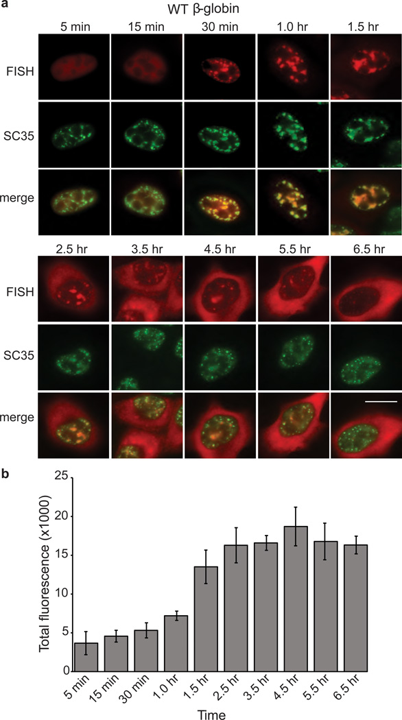
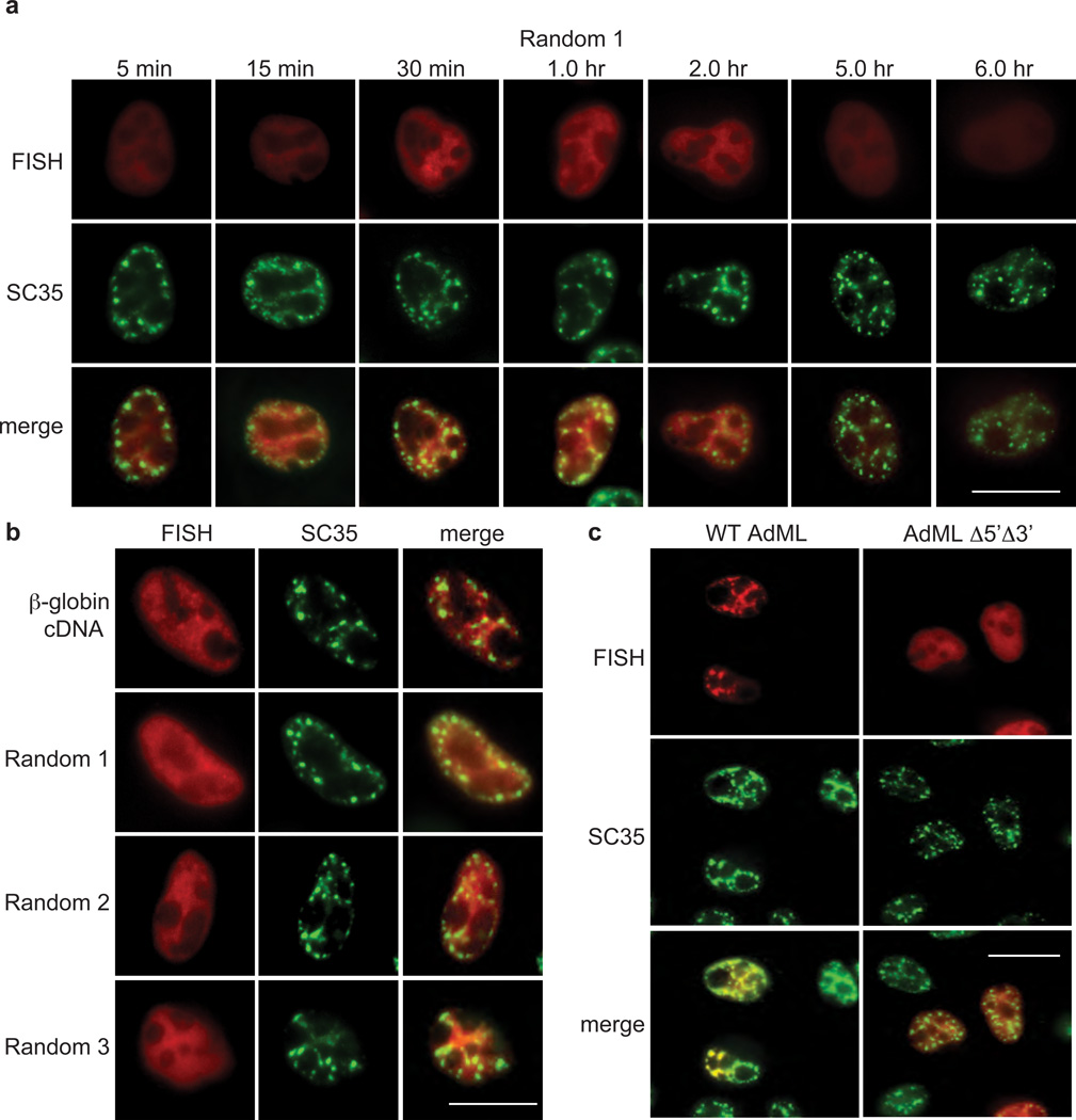
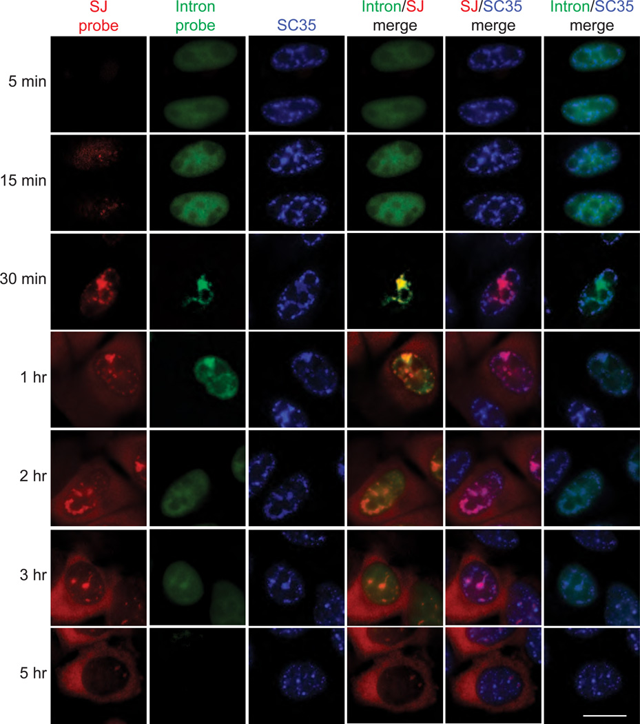
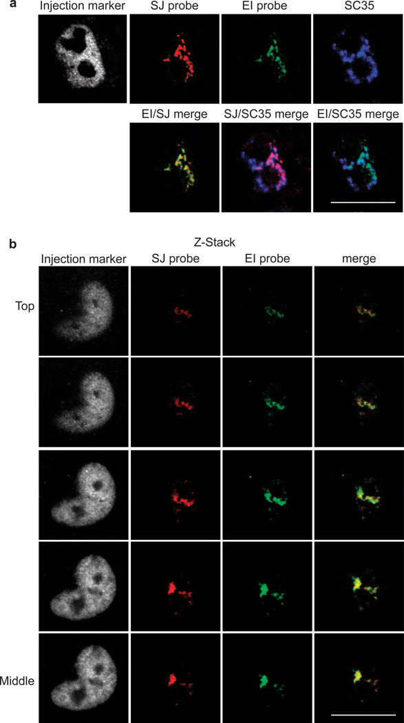
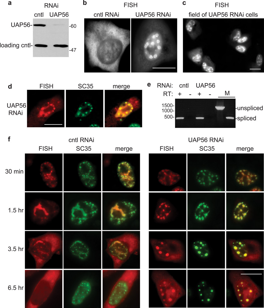
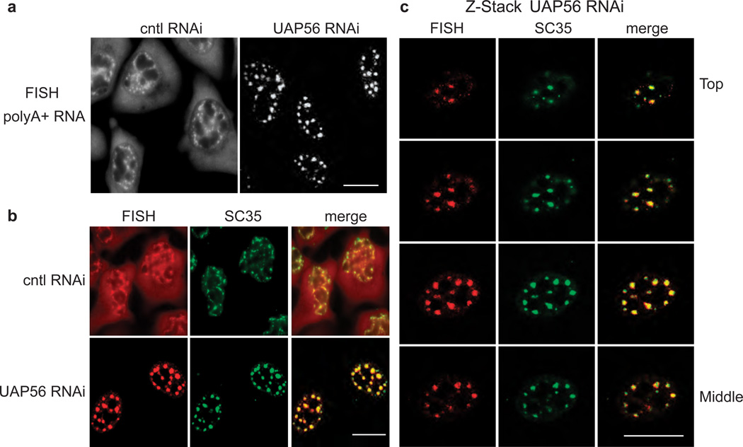
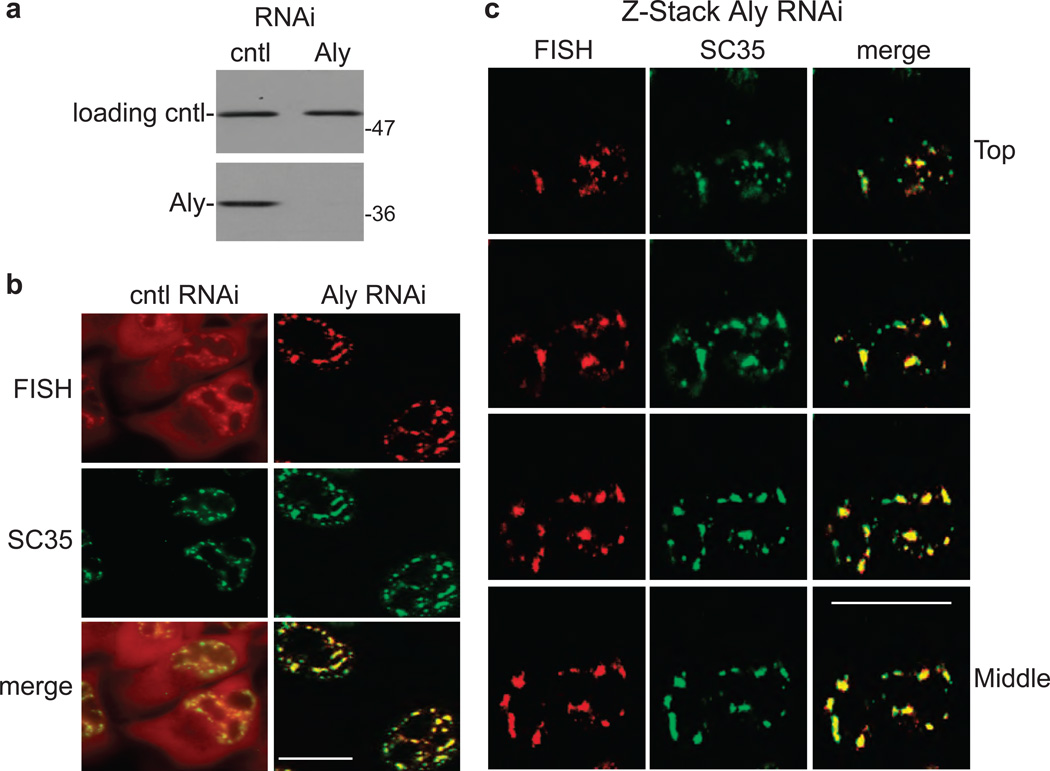
The TREX complex, which functions in mRNA export, is recruited to mRNA during splicing. Both the splicing machinery and the TREX complex are concentrated in 20-50 discrete foci known as nuclear speckle domains. In this study, we use a model system where DNA constructs are microinjected into HeLa cell nuclei, to follow the fates of the transcripts. We show that transcripts lacking functional splice sites, which are inefficiently exported, do not associate with nuclear speckle domains but are instead distributed throughout the nucleoplasm. In contrast, pre-mRNAs containing functional splice sites accumulate in nuclear speckles, and our data suggest that splicing occurs in these domains. When the TREX components UAP56 or Aly are knocked down, spliced mRNA, as well as total polyA+ RNA, accumulates in nuclear speckle domains. Together, our data raise the possibility that pre-mRNA undergoes splicing in nuclear speckle domains, before their release by TREX components for efficient export to the cytoplasm.


Figures

Figure 1
Figure 1. FISH of RNAs after transcription of CMV-DNA constructs
(a) Schematic of constructs. The CMV promoter and BGH polyA sites and the location of the FISH probe (indicated by short line) are shown. The sizes (in nts) of the introns, exons, cDNA and random transcripts are indicated for the respective constructs. (b) FISH of indicated transcripts detected 5 mins after microinjection of CMV-DNA constructs (50 ng/µl) into HeLa cell nuclei. FITC-conjugated dextran was co-injected as a nuclear marker (inset panels). Scale bar, 10 µm. (c) Quantification of total FISH fluorescence was determined for a minimum of 40 cells for each construct. Data represent the average of three experiments, and error bars indicate standard error.
Figure 2
Figure 2. Wild type β-globin transcripts associate with nuclear speckle domains
(a) FISH time course showing nucleocytoplasmic distribution of β-globin transcripts after microinjection of wild type (WT) β-globin DNA construct into HeLa cell nuclei. α- amanitin was added at the 30 min time point to block further transcription. Cells were incubated for the indicated times before fixation. FISH, IF using an SC35 monoclonal antibody, and merged images are indicated. Scale bar, 10 µm. (b) Quantification of total FISH fluorescence was determined for a minimum of 10 cells for each time point. Data represent the average of three experiments, and error bars indicate standard error.
Figure 3
Figure 3. Splice-site dependent association of transcripts with nuclear speckle domains
(a) CMV Random 1-DNA construct was microinjected in HeLa nuclei, and α-amanitin was added after 30 mins to block further transcription. A FISH time course for Random 1 RNA was carried out at the indicated times. FISH, IF using an SC35 monoclonal antibody, and merged images are shown. Scale bar, 10 µm. (b) same as (a) except Random 1, 2, and 3 CMV-DNA constructs and CMV β-globin cDNA-construct were microinjected, and FISH was carried out 1 hr after microinjection. (c) same as (a) except CMV-WT AdML or CMV-Δ5’ Δ3’ AdML DNA constructs were used, and FISH was carried out 30 mins after microinjection. FISH, IF using an SC35 monoclonal antibody, and merged images are shown. Scale bar, 10 µm.
Figure 4
Figure 4. FISH time course ofβ-globin transcripts using splice junction- or intron-specific probes
(a) CMV β-globin-DNA construct was microinjected into nuclei, α-amanitin was added at 30 mins, and FISH was carried out at the indicated times. Probes were to the second splice junction (SJ probe) or the second intron (intron probe) of β-globin mRNA. IF was carried out using an SC35 monoclonal antibody. The indicated merged FISH and IF images are shown. Scale bar, 10 µm.
Figure 5
Figure 5. Evidence thatβ-globin pre-mRNA is spliced in a subset of nuclear speckle domains
(a) CMV-β-globin DNA was microinjected into nuclei, and FISH was carried out after 30 mins using a probe to the splice junction (SJ probe) or the intron-exon junction (IE probe). IF with SC35 monoclonal antibody was carried out after the FISH. Confocal microscopy was used to visualize the cells. The nucleus was detected by the injection marker. The indicated merged images are shown in the second row. (b) Z-Stacked confocal images for a representative nucleus using the indicated probes or SC35 antibody for IF are indicated. Z-stacks were taken in steps of µm. The Top to Middle sections of the Z-stacks are shown. Scale bar, 10 µm.
Figure 6
Figure 6. β-globin mRNA accumulates in nuclear speckle domains in UAP56 knockdown cells
(a) Western analysis using whole cell lysates from control (cntl) or UAP56 knockdown cells probed with an antibody against UAP56 or an antibody against tubulin as a loading control. Molecular weight markers in kilodaltons are shown. (b) CMV-β-globin DNA construct was microinjected into the nuclei of control knockdown or UAP56 knockdown cells followed by FISH for β-globin mRNA. Scale bar, 10µm. (c) Same as (b), but shown at lower magnification. Scale bar, 10µm. (d) FISH for β-globin mRNA in UAP56 knockdown cells 3.5 hr after microinjection, SC35 IF using an SC35 monoclonal antibody, and merged FISH and IF images are shown. Scale bar, 10µm. (e) RT-PCR of total RNA extracted from control knockdown and UAP56 knockdown cells microinjected with β-globin DNA. RT (reverse transcriptase): + and − indicates reactions carried out in the presence or absence of RT, followed by PCR. DNA size markers (in base pairs) are indicated on the left. M: marker for unspliced and spliced mRNAs. (f) FISH time course showing nucleocytoplasmic distribution of β-globin transcripts after microinjection of wild type (WT) β-globin DNA construct into the nuclei of control knockdown or UAP56 knockdown cells. α-amanitin was added at the 30 min time point to block further transcription. Cells were incubated for the indicated times before fixation. FISH, IF using an SC35 monoclonal antibody, and merged FISH and IF images are shown. Scale bar, 10 µm.
Figure 7
Figure 7. PolyA+ RNA accumulates in nuclear speckle domains in UAP56 knockdown cells
(a) FISH of polyA+ RNA in control and UAP56 knockdown cells. Scale bar, 10µm. (b) FISH for polyA+ RNA followed by IF using an SC35 monoclonal antibody in control or UAP56 knockdown cells. The merged FISH and IF images are shown in the right panel. Scale bar, 10 µm. (c) Z-Stack confocal analysis of FISH for polyA+ RNA, SC35 IF, and merged images are indicated. Z-stacks were taken in steps of µm. The Top to Middle sections of the Z-stacks is shown. Scale bar, 10 µm.
Figure 8
Figure 8. PolyA+ RNA accumulates in nuclear speckle domains in Aly knockdown cells
(a) Western analysis using whole cell lysates from control (cntl) or Aly knockdown cells probed with an antibody against Aly or against tubulin as a loading control. Molecular weight markers in kilodaltons are shown. (b) FISH for polyA+ RNA followed by IF using an SC35 monoclonal antibody in control or Aly knockdown cells. The merged FISH and IF images are shown in the bottom panels. Scale bar, 10 µm. (c) Z-Stack confocal analysis of FISH for polyA+ RNA, SC35 IF, and merged images are indicated. Z-stacks were taken in steps of µm. The Top to Middle sections of the Z-stacks are shown. Scale bar, 10 µm

References

    1. Maniatis T, Reed R. An extensive network of coupling among gene expression machines. Nature. 2002;416:499–506. - PubMed
    1. Reed R. Coupling transcription, splicing and mRNA export. Curr Opin Cell Biol. 2003;15:326–331. - PubMed
    1. Pandit S, Wang D, Fu XD. Functional integration of transcriptional and RNA processing machineries. Curr Opin Cell Biol. 2008;20:260–265. - PMC - PubMed
    1. Zhong XY, Wang P, Han J, Rosenfeld MG, Fu XD. SR proteins in vertical integration of gene expression from transcription to RNA processing to translation. Mol Cell. 2009;35:1–10. - PMC - PubMed
    1. Hall LL, Smith KP, Byron M, Lawrence JB. Molecular anatomy of a speckle. Anat Rec A Discov Mol Cell Evol Biol. 2006;288:664–675. - PMC - PubMed

Publication types