. 2010 Oct 14:2010:179594.
doi: 10.4061/2010/179594.

Molecular mechanisms of the whole DNA repair system: a comparison of bacterial and eukaryotic systems

Affiliations

Molecular mechanisms of the whole DNA repair system: a comparison of bacterial and eukaryotic systems

Rihito Morita et al. J Nucleic Acids. .

Abstract

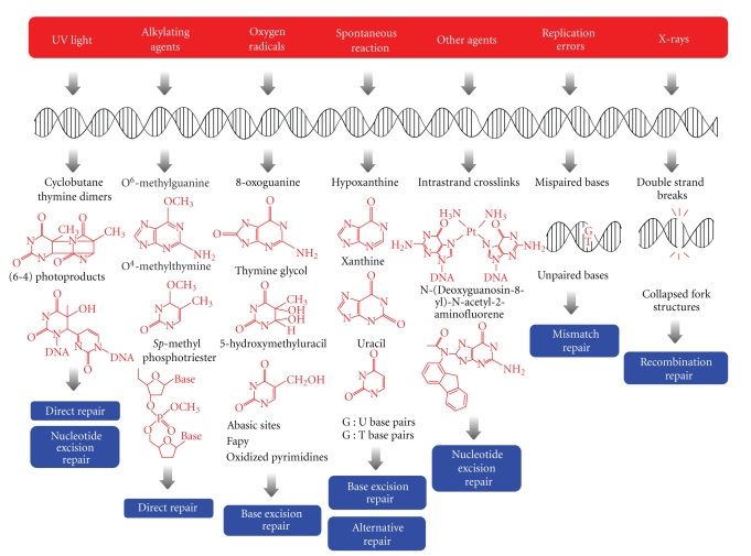
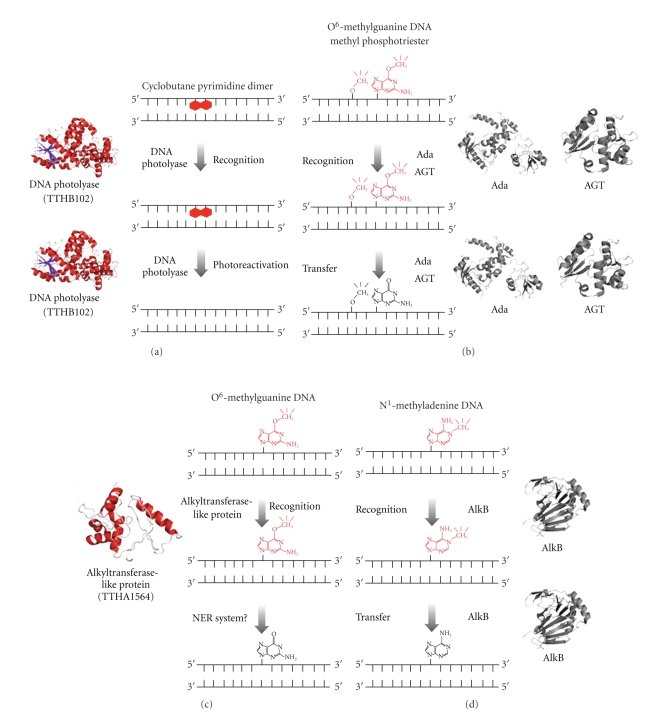
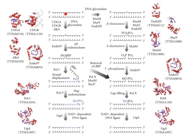
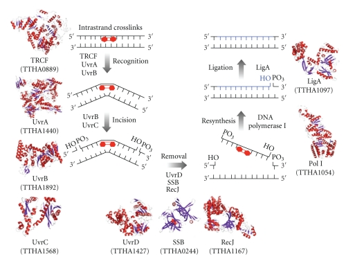
DNA is subjected to many endogenous and exogenous damages. All organisms have developed a complex network of DNA repair mechanisms. A variety of different DNA repair pathways have been reported: direct reversal, base excision repair, nucleotide excision repair, mismatch repair, and recombination repair pathways. Recent studies of the fundamental mechanisms for DNA repair processes have revealed a complexity beyond that initially expected, with inter- and intrapathway complementation as well as functional interactions between proteins involved in repair pathways. In this paper we give a broad overview of the whole DNA repair system and focus on the molecular basis of the repair machineries, particularly in Thermus thermophilus HB8.


Figures

Figure 1
Figure 1
Different repair systems for the principal types of DNA lesion produced by a wide range of factors. UV-light induces cyclobutane pyrimidine dimers or (6-4) photoproducts that are repaired by nucleotide excision repair and direct reversal systems. Alkylating agents can modify all of the bases and the phosphates of the DNA, and some repair proteins remove these alkyl adducts in a direct manner. Oxygen radicals modify DNA, and the base excision repair system acts to reverse these changes. The main cause of spontaneous mutation is deamination, and base excision repair and alternative repair systems remove the lesions. Other bulky adducts or interstrand cross-links are repaired by the nucleotide excision repair system. The mismatch repair pathway repairs replication errors. Double-strand breaks and four-way junctions are induced by X-rays and are repaired by recombinational repair.
Figure 2
Figure 2
A schematic representation of models for direct reversal of DNA damage. The structure of the ATL proteins was modeled by SWISS-MODEL (the template structure is Sulfolobus tokodaii Ogt) [19, 20]. AGT, Ada, and AlkB are not conserved in T. thermophilus. (a) Cyclobutane pyrimidine dimers are recognized by photolyase (TTHB102; PDB ID: 1IQR) and repaired by photolyase. (b) O 6-methylguanines are recognized by AGT (PDB ID: 1EH6) in most species and by the C-terminal domain of Ada (PDB ID: 1SFE) in E. coli. Methyl phosphotriesters are recognized by the N-terminal domain of Ada (PDB ID: 1WPK) in E. coli. These enzymes directly accept a methyl group, and the alkyl adducts are removed from the DNA. (c) O 6-alkyl adducts including O 6-methylguanines are recognized by ATL proteins (TTHA1564; predicted model) in several species. It is predicted that NER proteins are involved in this pathway after recognition of the adducts by ATL proteins. (d) N 1-methyladenines and N 3-methylcytosines are recognized by AlkB (PDB ID: 2IUW). Methyl group transfer by AlkB depends on α-ketoglutarate and Fe(II).
Figure 3
Figure 3
General mechanism of the BER pathway in T. thermophilus. UDGA, UDGB, and AlkA are monofunctional DNA glycosylases. UDGA (PDB ID: 1UI0) and UDGB (PDB ID: 2DDG) remove uracil from DNA. AlkA removes 3-methyladenine in E. coli. MutY and EndoIII are bifunctional DNA glycosylases and have both DNA glycosylase and AP lyase activities. MutY removes adenine opposite 8-oxoG, and EndoIII removes pyrimidine residues damaged by ring saturation, fragmentation, and contraction [41], by which 3′-phospho α,β-unsaturated aldehyde (3′-PUA) remains. MutM (PDB ID: 1EE8) is a trifunctional DNA glycosylase that removes 8-oxoG from oxidatively damaged DNA and 3′-phosphate remains. An AP site resulting from DNA glycosylase activity is processed by EndoIV or multifunctional DNA glycosylases. EndoIV has both AP endonuclease activity and 3′-esterase activity in E. coli [42, 43]. PolX or MutM removes 5′-dRP by dRP lyase activity. In addition, 5′-3′ exonuclease (RecJ) may have dRPase activity. The resulting gap is filled by PolI or PolX followed by sealing of the nick by LigA. The structures of AlkA, EndoIII, MutY, PolI, PolX, and LigA were obtained using SWISS-MODEL [19, 20] (PDB ID: 2H56, 2ABK, 3FSP, 1TAU, 2W9M, and 1V9P, resp.) based on amino acids sequences of T. thermophilus HB8.
Figure 4
Figure 4
A schematic representation of models for the nucleotide excision repair pathway controlled by Uvr proteins. All of the predicted protein structures were modeled using SWISS-MODEL. The template structures used in the model building were Geobacillus stearothermophilus UvrA, the N- and C-terminal domain of Thermotoga maritime UvrC, G. stearothermophilus UvrD, Thermus aquaticus DNA polymerase I, Thermus filiformis DNA ligase, and E. coli TRCF. UvrA (TTHA1440; predicted model) and UvrB (TTHA1892; PDB ID: 1D2M) recognize the DNA lesion. In transcribing strand, TRCF (TTHA0889; predicted model) is also involved in recognition of the lesion. UvrC (TTHA1568; predicted model) incises both sides of the lesion. The DNA fragment containing the lesion is excised by UvrD (TTHA1427; predicted model), SSB (TTHA0244; 2CWA), and exonuclease RecJ (TTHA1167; PDB ID: 2ZXO). A new strand is resynthesized by DNA polymerase I (TTHA1054; predicted model) and ligated by DNA ligase (TTHA1097; predicted model).
Figure 5
Figure 5
The domain architectures of UvrB, UvrA, and TRCF. (a) UvrB is comprised of five domains. Domains 1a (yellow) and 3 (red) contain helicase motifs. Domain 1b (green) has the flexible β-hairpin invosved in substrate recognition. Domain 2 (blue) interacts with UvrA. Domain 4 is disordered in the crystal structures. (b) UvrA is comprised of six domains: ATP-binding I (red), ATP-binding II (blue), signature I (pink), signature II (cyan), UvrB-binding (yellow), and insertion (green) domain. The white region is the other subunit of the dimer. (c) TRCF is comprised of seven domains. Domains 1 and 2 (not separated in the figure) comprise UvrB homology module (blue). Domains 5 (yellow) and 6 (green) comprise DNA translocation module. Relay helix (yellow) interacts with domain 4 (pink), RNA polymerase interaction domain (RID). The functions of domain 3 (orange) and domain 7 (red) are unclear.
Figure 6
Figure 6
A schematic representation of models for MMR pathways in E. coli and mutH-less bacteria. (a) 5′- and 3′-methyl-directed MMR in E. coli. DNA mismatches principally result from misincorporation of bases during DNA replication. The MutS (PDB ID: 1E3M)/MutL (PDB ID: 1NHJ) complex recognizes a mismatch and activates the MutH endonuclease (PDB ID: 1AZO). MutH nicks the unmethylated strand of the duplex to introduce an entry point for the excision reaction. In 3′-methyl-directed MMR, one of the 5′-3′ exonucleases (RecJ and exonuclease VII (ExoVII)) removes the error-containing DNA strand in cooperation with UvrD helicase (PDB ID: 2IS4) and single-stranded DNA-binding protein (SSB; PDB ID: 1EYG). By contrast, one of the 3′-5′ exonucleases (exonuclease I (ExoI; PDB ID: 1FXX) and exonuclease X (ExoX)) is responsible for the 3′-5′ excision reaction. DNA polymerase III (PDB ID: 2HNH) and DNA ligase (PDB ID: 2OWO) synthesize a new strand to complete the repair. (b) A predicted model for 5′- and 3′-nick-directed MMR in T. thermophilus. After recognition of a mismatch by MutS (TTHA1324), MutL (TTHA1323) incises the discontinuous strand of the mismatched duplex to direct the excision reaction to the newly synthesized strand. The error-containing DNA segment is excised by UvrD helicase (TTHA1427), SSB (TTHA0244), and an exonuclease (either RecJ (TTHA1167; PDB ID: 2ZXR) or ExoI (TTHB178)) followed by the resynthesis of a new strand by DNA polymerase III (TTHA0180) and DNA ligase (TTHA1097). The modeled structures of T. thermophilus MutS, MutL (amino acid residues 1–316), ExoI, DNA polymerase III α subunit, DNA ligase, and E. coli RecJ were modeled using SWISS-MODEL. The template structures used for model building were E. coli MutS, the N-terminal domain of MutL, ExoI, UvrD, DNA polymerase III α subunit, DNA ligase, and T. thermophilus RecJ.
Figure 7
Figure 7
A schematic pathway of recombination repair and structures of the proteins involved in T. thermophilus. Recombination repair of DSBs is initiated by an end resection step in which DSB ends are processed by the concerted action of RecJ nuclease (TTHA1167; PDB ID: 2ZXR) and SSB (TTHA0244; PDB ID: 2CWA) to form 3′-ssDNA tails. After end resection, the SSB-ssDNA complex is disassembled and RecA recombinase (TTHA1818) is loaded onto ssDNA by “mediators”, RecF (TTHA0264), RecO (TTHA0623), and RecR (TTHA1600), to promote strand invasion. DNA repair synthesis is primed by PolI (TTHA1054) and PolIII (TTHA0180) from the invaded strand of the D-loop structure. Alternatively, second-end capture is mediated by RecO and SSB and branch migration mediated by the RuvA-RuvB complex (TTHA0291-TTHA0406; PDB ID: 1IXR) and RecG (TTHA1266) to yield HJs. HJs are cleaved by RuvC resolvase (TTHA1090) and the nicks sealed by LigA (TTHA1097). Newly synthesized DNA is colored in blue. The model structures of T. thermophilus RecA, RecF, RecO, RecR, PolI, PolIII α subunit, RecG, RuvC, and LigA were generated using SWISS-MODEL. The models were based on the structures of Mycobacterium smegmatis RecA (PDB ID: 2OE2), D. radiodurans RecF (PDB ID: 2O5V), RecO (PDB ID: 1U5K), RecR (PDB ID: 1VDD), E. coli PolI (PDB ID: 1TAU), PolIII α subunit (PDB ID: 2HNH), RuvC (PDB ID: 1HJR), LigA (PDB ID: 2OWO), and Thermotoga maritima RecG (PDB ID: 1GM5).
Figure 8
Figure 8
A schematic illustration of RecA-ssDNA interaction in the nucleoprotein filament. (a) A schematic representation of a RecA-ssDNA nucleoprotein filament. The filament comprises a helical structure. RecA molecules are shown as red spheres and the ssDNA as a black line. (b) A schematic model of RecA-ssDNA interaction. The RecA protomer has the L1 and L2 loops and the N-terminal region to make contact with the ssDNA. The bound ssDNA comprises a nucleotide triplet with a nearly normal B-form distance between bases followed by a long internucleotide stretch before the next triplet. The ATP binds to RecA-RecA interfaces. The schematic model was prepared from the crystal structure of RecA-ssDNA complex (PDB ID: 3CMW).

References

    1. Schärer OD. Chemistry and biology of DNA repair. Angewandte Chemie. 2003;42(26):2946–2974. - PubMed
    1. Altieri F, Grillo C, Maceroni M, Chichiarelli S. DNA damage and repair: from molecular mechanisms to health implications. Antioxidants and Redox Signaling. 2008;10(5):891–937. - PubMed
    1. Friedberg EC, Walker GC, Siede W. DNA Repair and Mutagenesis. Washington, DC, USA: ASM Press; 2006.
    1. Oshima T, Imahori K. Description of Thermus thermophilus (Yoshida and Oshima) comb. nov., a nonsporulating thermophilic bacterium from a Japanese thermal spa. International Journal of Systematic Bacteriology. 1974;24(1):102–112.
    1. Iino H, Naitow H, Nakamura Y, et al. Crystallization screening test for the whole-cell project on Thermus thermophilus HB8. Acta Crystallographica Section F. 2008;64(6):487–491. - PMC - PubMed

LinkOut - more resources