Giant adrenal myelolipoma: Incidentaloma with a rare incidental association
- PMID: 20981204
- PMCID: PMC2955231
- DOI: 10.4103/0974-7796.68865
Giant adrenal myelolipoma: Incidentaloma with a rare incidental association
Abstract
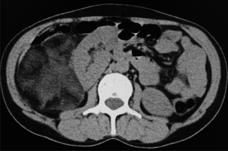
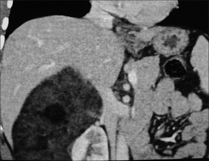
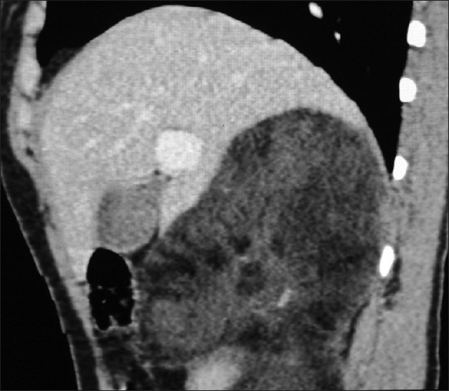
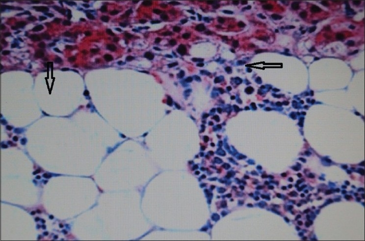
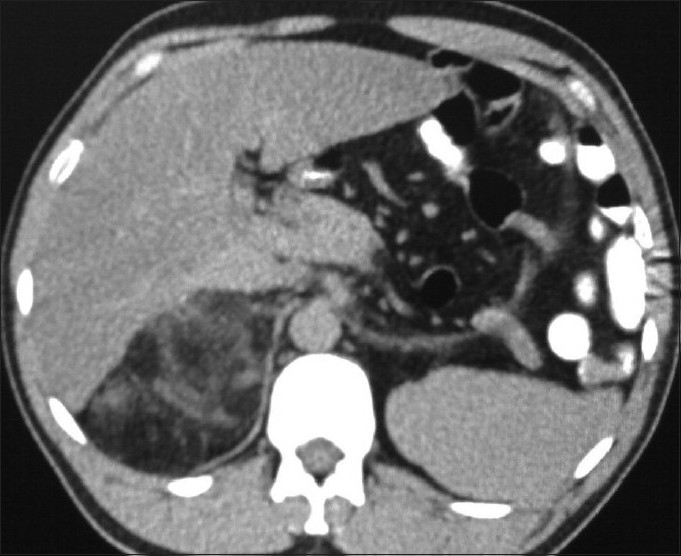
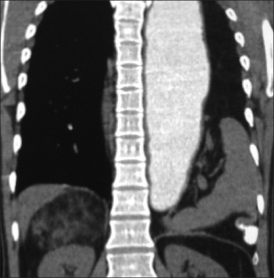
Adrenal myelolipoma is an unusual, benign and biochemically inactive tumor that is composed of mature adipose and hematopoietic tissue. It is usually diagnosed accidentally and nowadays much more frequently because of widespread use of ultrasonography, computed tomography (CT) and magnetic resonance imaging. Adrenal myelolipoma is usually unilateral and asymptomatic, though known to be associated with obesity, hypertension, endocrinological disorders and some malignancies. We report herein two cases of right-sided giant adrenal myelolipoma diagnosed by multidetector-row CT. One patient was symptomatic because of a large mass in the right upper abdomen, which on imaging with CT was seen to be right adrenal myelolipoma. Another patient had a large left side Bochdalek hernia and right adrenal myelolipoma was incidentally discovered on CT.
Keywords: Adrenal gland; Bochdalek hernia; multidetector-row CT; myelolipoma.
Conflict of interest statement
Figures
References
-
- Wagnerová H, Lazúrová I, Bober J, Sokol L, Zachar M. Adrenal myelolipoma. 6 cases and a review of the literature. Neoplasma. 2004;51:300–5. - PubMed
-
- Oberling C. Les formations myelolipomateuses. Bull Assoc Fr Etud Cancer. 1929;18:234–46.
-
- Gierke E. Uber Knochenmarksgwebe in der Nebenniere. Zeiglers Beitr Path Anat. 1905;7:311–24.
-
- Yildiz L, Akpolat I, Erzurumlu K, Aydin O, Kandemir B. Giant adrenal myelolipoma: case report and review of the literature. Pathol Int. 2000;50:502–4. - PubMed
-
- Daneshmand S, Quek ML. Adrenal myelolipoma: diagnosis and management. J Urol. 2006;3:71–4. - PubMed