. 2010 Jan;1(1):26-39.
doi: 10.1177/1947601909358184.

Germinal Cell Aplasia in Kif18a Mutant Male Mice Due to Impaired Chromosome Congression and Dysregulated BubR1 and CENP-E

Affiliations

Germinal Cell Aplasia in Kif18a Mutant Male Mice Due to Impaired Chromosome Congression and Dysregulated BubR1 and CENP-E

Xue-Song Liu et al. Genes Cancer. 2010 Jan.

Abstract

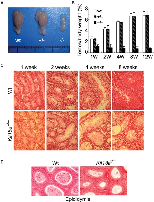
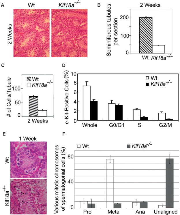
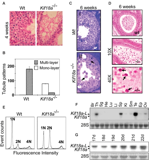
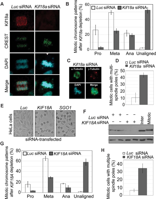
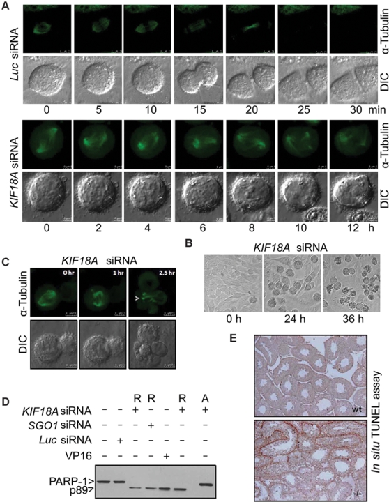
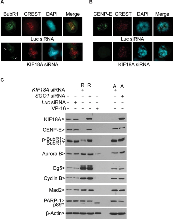
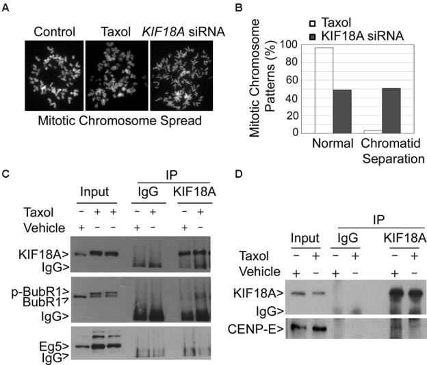
Chromosomal instability during cell division frequently causes cell death or malignant transformation. Orderly chromosome congression at the metaphase plate, a paramount process to vertebrate mitosis and meiosis, is controlled by a number of molecular regulators, including kinesins. Kinesin-8 (Kif18A) functions to control mitotic chromosome alignment at the mid-zone by negative regulation of kinetochore oscillation. Here the authors report that disrupting Kif18a function results in complete sterility in male but not in female mice. Histological examination reveals that Kif18a(-/-) testes exhibit severe developmental impairment of seminiferous tubules. Testis atrophy in Kif18a(-/-) mice is caused by perturbation of microtubule dynamics and spindle pole integrity, leading to chromosome congression defects during mitosis and meiosis. Depletion of KIF18A via RNAi causes mitotic arrest accompanied by unaligned chromosomes and increased microtubule nucleating centers in both GC-1 and HeLa cells. Prolonged depletion of KIF18A causes apoptosis due to perturbed microtubule dynamics. Further studies reveal that KIF18A silencing results in degradation of CENP-E and BubR1, which is accompanied by premature sister chromatid separation. KIF18A physically interacts with BubR1 and CENP-E, and this interaction is modulated during mitosis. Combined, the studies indicate that KIF18A is essential for normal chromosome congression during cell division and that the absence of KIF18A function causes severe defects in microtubule dynamics, spindle integrity, and checkpoint activation, leading to germinal cell aplasia in mice.

Keywords: BubR1; KIF18A; chromosome congression; knockout mice; testis development.


Conflict of interest statement

The authors declared no potential conflicts of interest with respect to the authorship and/or publication of this article.

Figures

Figure 1.
Figure 1.
Disruption of Kif18a causes testis atrophy and germ cell aplasia. (A) Morphologies of representative testes from 8-week-old mice of various genotypes. (B) Testes of individual mice, as well as their bodies, of various genotypes were weighed during various stages of development (weeks 1, 2, 4, 8, and 12). The testes/body weight ratios were calculated for wild-type mice (wt), heterozygotes (+/−), and homozygotes (−/−). (C) Sections of wt and Kif18a−/− testes at different developmental stages were stained with H&E. Representative images are shown. (D) Sections of wt and Kif18a−/− epididymis at week 4 were stained with H&E, and representative images are shown.
Figure 2.
Figure 2.
Mitotic defects in Kif18a−/− mouse testes. (A) Representative sections of wt and Kif18a−/− at week 2 were stained with H&E. (B) Seminiferous tubules were counted from 6 random fields of each wt or Kif18a−/− testis. The data were summarized from 3 independent mice. (C) Cells in each seminiferous wt or Kif18a−/− tubule were counted from 6 random fields of each testis. The data were summarized from 3 independent mice. (D) The c-Kit-positive cells isolated from wt or Kif18a−/− testes were subjected to the DNA content analysis by flow cytometry. Representative results are shown. (E) Representative wt and Kif18a−/− seminiferous tubules at week 1 are presented. Arrowheads denote mitotic cells with unaligned chromosomes. (F) Mitotic cells in week 1 wt or Kif18a−/− seminiferous tubules were examined under microscope. Cells of various mitotic stages or with unaligned chromosomes were quantified.
Figure 3.
Figure 3.
Meiotic defects in Kif18a−/− mouse testes and tissue-specific expression of Kif18a. (A) Wt and Kif18a−/− seminiferous tubules at week 4 were stained with H&E. Representative tubules are shown. (B) Seminiferous tubules with monolayer or multilayers of cells were counted from wt or Kif18a−/− testes. The data were summarized from 6 individual testes. (C) H&E-stained wt (I) and Kif18a−/− (II) seminiferous tubules at week 6. Arrows denote the spermatids with multiple or bi-nuclei. (D) H&E-stained wt and Kif18a−/− epididymis. Arrows denote the nonmotile bi-nucleated spermatozoa. (E) Testicular cells isolated from wt and Kif18a−/− testes at week 6 were stained with propidium iodide and subjected to DNA content analysis by flow cytometry. (F) Equal amounts of total RNA isolated from various mouse organs were subjected to Northern blotting analysis using Kif18a cDNA as a probe. Br, Th, Sg, He, Lu, Li, Sp, Ki, Sm, Te, Ep, and Ov stand for brain, thymus, salivary glands, heart, lung, liver, spleen, kidney, small intestine, testes, epididymis, and ovaries, respectively. (G) Equal amounts of total RNA isolated from wild-type testes of various developmental stages (days 17-22) were subjected to Northern blotting using Kif18a cDNA as a probe. Kif18a-L stands for the large form of Kif18a transcript.
Figure 4.
Figure 4.
KIF18A depletion causes spindle pole and chromosome congression defects in vitro. (A) Mouse spermatogonial GC-1 cells transfected with Kif18a or luciferase (Luc) siRNA for 24 h were fixed and stained with antibodies to KIF18A (green) and CREST (red). DNA was stained with 4′6′-diamidino-2-phenylindole (DAPI; blue). Representative results are shown. (B) Cells at various mitotic stages or with unaligned/misaligned chromosomes were summarized from GC-1 cells transfected with Kif18a or luciferase siRNA. The data were summarized from 3 independent experiments. (C) GC-1 cells transfected with Kif18a or luciferase siRNA for 24 h were fixed and stained with antibodies to α-tubulin (green) and γ-tubulin (red). DNA was stained with DAPI (blue). Representative images are shown. (D) GC-1 cells transfected with Kif18a or luciferase siRNA for 24 h were fixed and stained with antibodies to α-tubulin (green) and γ-tubulin (red). Mitotic cells with multiple spindle poles were summarized from 3 independent experiments. (E) HeLa cells transfected with KIF18A or luciferase siRNA for 24 h were examined under a light microscope. Representative images are shown. (F) Equal amounts of proteins from HeLa cells transfected with KIF18A or luciferase siRNA for 24 h were blotted for KIF18A and β-actin. (G) Mitotic cells of various stages or with unaligned/misaligned chromosomes were counted from HeLa cells transfected with KIF18A or luciferase siRNA for 24 h. The data were summarized from 3 independent experiments. (H) HeLa cells transfected with KIF18A or luciferase siRNA for 24 h were fixed and stained with antibodies to α-tubulin (green) and γ-tubulin (red). Mitotic cells with multiple spindle poles were summarized from 3 independent experiments.
Figure 5.
Figure 5.
KIF18A depletion induces prolonged mitotic arrest, leading to mitotic catastrophe. (A) HeLa cells constitutively expressing green fluorescent protein (GFP)–tubulin were transfected with KIF18A or luciferase siRNA for 24 h. These cells were then subjected to time lapse confocal microscopy analysis. Representative differential interference contrast (DIC) and α-tubulin images of metaphase cells undergoing mitosis are shown. (B) HeLa cells transfected with KIF18A or luciferase siRNA for 0, 24, or 36 h were observed under a light microscope. Representative images are shown. (C) HeLa cells constitutively expressing GFP-tubulin were transfected with KIF18A siRNA for 36 h. Mitotic cells were then subjected to time lapse confocal microcopy. DIC and α-tubulin images of a representative cell undergoing mitosis are shown. Arrows indicate the distorted, bundled microtubules. (D) HeLa cells transfected with KIF18A, SGO1, or luciferase siRNA for 24 h were collected and lysed. HeLa cells treated with VP16 overnight were also used for lysate preparation. Equal amounts of proteins were blotted for PARP-1 or its degradation product p89. R and A denote rounded-up cells and adherent cells, respectively. (E) Sections of wt and Kif18a−/− seminiferous tubules were subjected to terminal deoxynucleotidyl transferase–mediated dUTP-biotin nick end labeling (TUNEL) analysis. Representative images are shown.
Figure 6.
Figure 6.
KIF18A regulates the stability of BubR1 and CENP-E during mitosis. (A) HeLa cells transfected with KIF18A or luciferase siRNA for 24 h were fixed and stained with antibodies to α-BubR1 (green) and CREST (red). DNA was stained with 4′6′-diamidino-2-phenylindole (DAPI; blue). Representative images are shown. Arrows indicate BubR1 signals on unaligned chromosomes. (B) HeLa cells transfected with KIF18A or luciferase siRNA for 24 h were fixed and stained with antibodies to CENP-E (green) and CREST (red). DNA was stained with DAPI (blue). Representative images are shown. (C) HeLa cells transfected with KIF18A, SGO1, or luciferase siRNA for 24 h were collected and lysed. HeLa cells treated with VP16 overnight were also used for lysate preparation. Equal amounts of proteins from various treatments were blotted for KIF18A, CENP-E, BubR1, Aurora B, Eg5, cyclin B, Mad2, PARP-1, and β-actin. R and A denote rounded-up cells and adherent cells, respectively. Arrow p-BubR1 denotes the phosphorylated BubR1. R and A denote rounded-up and adherent cells, respectively.
Figure 7.
Figure 7.
KIF18A physically interacts with BubR1 and CENP-E. (A) Control HeLa cells or the cells treated with paclitaxel or transfected with KIF18A siRNA for 24 h were subjected to chromosome spread analysis. Representative mitotic chromosomes from each treatment are shown. (B) HeLa cells treated with paclitaxel or transfected with KIF18A siRNA for 24 h were subjected to chromosome spread analysis. The percentages of normal chromosomes and chromosomes with premature sister chromatid separation were summarized from 3 independent experiments. (C) Equal amounts of interphase cell or mitotic cell (Taxol-treated) lysates were subjected to immunoprecipitation using the KIF18A antibody or a control IgG. Immunoprecipitates, along with the lysate inputs, were blotted for KIF18A, BubR1, and Eg5. Arrow p-BubR1 denotes the phosphorylated BubR1. (D) Equal amounts of interphase cell or mitotic cell (Taxol-treated) lysates were subjected to immunoprecipitation using the KIF18A antibody or a control IgG. Immunoprecipitates and the lysate inputs were blotted for KIF18A and CENP-E. Each experiment was repeated for at least 3 times. Representative data are shown.

References

    1. Matzuk MM, Lamb DJ. Genetic dissection of mammalian fertility pathways. Nat Cell Biol 2002;4(suppl):s41-9 - PubMed
    1. Matzuk MM, Lamb DJ. The biology of infertility: research advances and clinical challenges. Nat Med 2008;14:1197-213 - PMC - PubMed
    1. de Kretser DM. Male infertility. Lancet 1997;349:787-90 - PubMed
    1. Meschede D, Horst J. The molecular genetics of male infertility. Mol Hum Reprod 1997;3:419-30 - PubMed
    1. Kulczycki LL, Kostuch M, Bellanti JA. A clinical perspective of cystic fibrosis and new genetic findings: relationship of CFTR mutations to genotype-phenotype manifestations. Am J Med Genet 2003;116A:262-7 - PubMed

LinkOut - more resources