. 2012 Sep;11(3):681-93.
doi: 10.1007/s12311-010-0224-3.

Controlling Ca2+-activated K+ channels with models of Ca2+ buffering in Purkinje cells

Affiliations

Controlling Ca2+-activated K+ channels with models of Ca2+ buffering in Purkinje cells

Haroon Anwar et al. Cerebellum. 2012 Sep.

Abstract

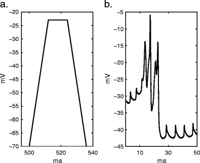
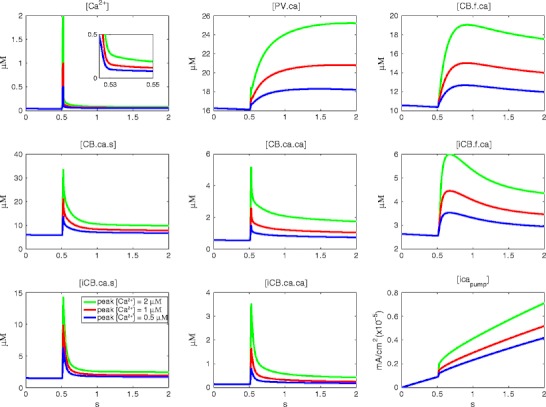
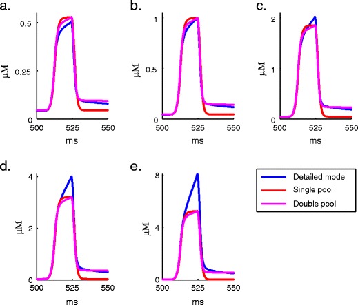
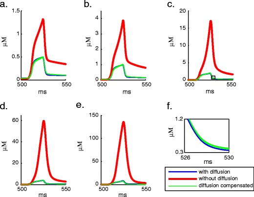
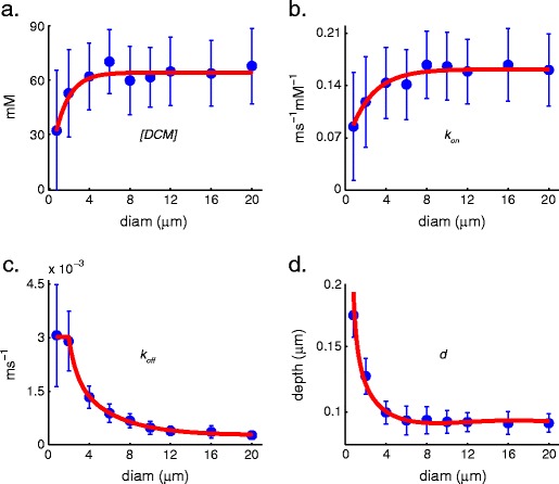
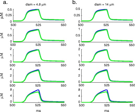
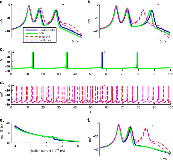
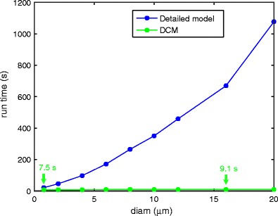
Intracellular Ca(2+) concentrations play a crucial role in the physiological interaction between Ca(2+) channels and Ca(2+)-activated K(+) channels. The commonly used model, a Ca(2+) pool with a short relaxation time, fails to simulate interactions occurring at multiple time scales. On the other hand, detailed computational models including various Ca(2+) buffers and pumps can result in large computational cost due to radial diffusion in large compartments, which may be undesirable when simulating morphologically detailed Purkinje cell models. We present a method using a compensating mechanism to replace radial diffusion and compared the dynamics of different Ca(2+) buffering models during generation of a dendritic Ca(2+) spike in a single compartment model of a PC dendritic segment with Ca(2+) channels of P- and T-type and Ca(2+)-activated K(+) channels of BK- and SK-type. The Ca(2+) dynamics models used are (1) a single Ca(2+) pool; (2) two Ca(2+) pools, respectively, for the fast and slow transients; (3) detailed Ca(2+) dynamics with buffers, pump, and diffusion; and (4) detailed Ca(2+) dynamics with buffers, pump, and diffusion compensation. Our results show that detailed Ca(2+) dynamics models have significantly better control over Ca(2+)-activated K(+) channels and lead to physiologically more realistic simulations of Ca(2+) spikes and bursting. Furthermore, the compensating mechanism largely eliminates the effect of removing diffusion from the model on Ca(2+) dynamics over multiple time scales.


Figures

Fig. 1
Fig. 1
a The depolarization command used to estimate Ca2+ profiles during generation of Ca2+ spikes. The peak voltage −22 mV was computed by taking an average of membrane potential during Ca2+ spikes (as shown in b), and the time of peak voltage ~12 ms was estimated from their average duration. b An example of electrophysiological dendritic Ca2+ spikes (provided by Ede Rancz and Michael Häusser, UCL, UK). Note that the small spikelets caused by retrograde conduction of somatic action potentials are not modeled in this study
Fig. 2
Fig. 2
Intracellular Ca2+ profiles simulated using the depolarization command shown in Fig. 1a, for different amplitudes of Ca2+ influx (indicated by color). The panels show, respectively, Ca2+ concentration ([Ca2+]), Ca2+ bound to PV (PV.ca), Ca2+ bound to slow binding site of CB (CB.f.ca), Ca2+ bound to fast binding site of CB (CB.ca.s), Ca2+ bound to fast and slow binding sites (CB.ca.ca), Ca2+ bound to slow binding site of immobile CB (iCB.f.ca), Ca2+ bound to fast binding site of immobile CB (iCB.ca.s), Ca2+ bound to fast and slow binding sites (iCB.ca.ca), and pump current over a period of 2,000 ms
Fig. 3
Fig. 3
Comparison of Ca2+ profiles generated with a voltage step protocol using single pool, double pool (parameters specified in the text), and detailed dynamics model. Different Ca2+ concentration peak amplitudes of a 0.5, b 1, c 2, d 4, and e 8 μM were simulated to demonstrate the problems the single pool or double pool models have in capturing the complex dynamics of the detailed model. See text for parameters of the pool models
Fig. 4
Fig. 4
Comparison of Ca2+ profiles of different peak amplitudes, a 0.5, b 1, c 2, d 4, and e 8 μM, simulated in a compartment with a diameter of 2 μm, using the detailed Ca2+ dynamics model, detailed Ca2+ dynamics model without diffusion, and DCM model. The removal of diffusion from the detailed model resulted in a steep rise in Ca2+, which could be well compensated by DCM. The inset (f) below the panel (c) highlights Ca2+ profiles simulated using five different sets of optimal DCM parameters found by using Neurofitter
Fig. 5
Fig. 5
Distribution of estimated DCM parameters ([DCM], k on, k off, and d; mean ± STD, n = 25: the five best solutions from five runs with different seeds for random number generation) for diffusion compensation plotted against compartment diameter. These were fitted respectively by a exponential function, b exponential function, c double exponential function, and d fifth order polynomial function
Fig. 6
Fig. 6
Comparison of Ca2+ profiles of different peak amplitudes simulated using detailed Ca2+ dynamics (blue) and diffusion compensated Ca2+ dynamics (green) with predicted values of parameters for diffusion compensation mechanism from the functions in Fig. 5. a Diameter 4.8 μm. b Diameter 14 μm
Fig. 7
Fig. 7
Dendritic Ca2+ spikes generated using different Ca2+ buffering models (aligned at the peak of the first spikelet in a, b, and f). The parameters of pool-based models are those used in Supplementary Fig. 1. The conductance values used to generate these spikes are listed in Table 2. a First burst of Ca2+ spikes. b Burst of Ca2+ spikes around 57 s. c, d Spontaneous Ca2+ spike bursting over 100 s: black asterisk indicates the first burst of Ca2+ spikes (shown in a), red asterisk indicates the burst of Ca2+ spikes around 57 s (shown in b). e Inter-burst interval (IBI) as a function of current injection. f Burst of Ca2+ spikes around 57 s with injection of 0.004 pA current
Fig. 8
Fig. 8
Comparison of run time for NEURON simulations of 10 s, with detailed Ca2+ dynamics and with DCM using compartments with different diameters. All the simulations were run on an Apple MacBook Pro, Intel Core 2 Duo 2.33 GHz

References

    1. Gähwiler BH, Llano I. Sodium and potassium conductances in somatic membranes of rat Purkinje cells from organotypic cerebellar cultures. J Physiol. 1989;417:105–22. - PMC - PubMed
    1. Gruol DL. Chronic exposure to alcohol during development alters the membrane properties of cerebellar Purkinje neurons in culture. Brain Res. 1991;558(1):1–12. doi: 10.1016/0006-8993(91)90707-3. - DOI - PubMed
    1. Knaus HG, Schwarzer C, Koch RO, Eberhart A, Kaczorowski GJ, Glossmann H, et al. Distribution of high-conductance Ca(2+)-activated K+ channels in rat brain: targeting to axons and nerve terminals. J Neurosci. 1996;16(3):955–63. - PMC - PubMed
    1. Jacquin TD, Gruol DL. Ca2+ regulation of a large conductance K+ channel in cultured rat cerebellar Purkinje neurons. Eur J Neurosci. 1999;11(2):735–9. doi: 10.1046/j.1460-9568.1999.00478.x. - DOI - PubMed
    1. Womack MD, Khodakhah K. Characterization of large conductance Ca2+-activated K+ channels in cerebellar Purkinje neurons. Eur J Neurosci. 2002;16(7):1214–22. doi: 10.1046/j.1460-9568.2002.02171.x. - DOI - PubMed

Publication types

MeSH terms

Substances

LinkOut - more resources