. 2011 Mar;21(2):215-24.
doi: 10.1111/j.1750-3639.2010.00441.x. Epub 2010 Oct 4.

Mutation analysis of CCM1, CCM2 and CCM3 genes in a cohort of Italian patients with cerebral cavernous malformation

Affiliations

Mutation analysis of CCM1, CCM2 and CCM3 genes in a cohort of Italian patients with cerebral cavernous malformation

Rosalia D'Angelo et al. Brain Pathol. 2011 Mar.

Erratum in

  • Brain Pathol. 2011 May;21(3):360

Abstract

Cerebral cavernous malformations (CCMs) are vascular lesions of the CNS characterized by abnormally enlarged capillary cavities. CCMs can occur as sporadic or familial autosomal dominant form. Familial cases are associated with mutations in CCM1[K-Rev interaction trapped 1 (KRIT1)], CCM2 (MGC4607) and CCM3 (PDCD10) genes. In this study, a three-gene mutation screening was performed by direct exon sequencing, in a cohort of 95 Italian patients either sporadic or familial, as well as on their at-risk relatives. Sixteen mutations in 16 unrelated CCM patients were identified,nine mutations are novel: c.413T > C; c.601C > T; c.846 + 2T > G; c.1254delA; c.1255-4delGTA; c.1682-1683 delTA in CCM1; c.48A > G; c.82-83dupAG in CCM2; and c.395 + 1G > A in CCM3 genes [corrected].The samples, negative to direct exon sequencing, were investigated by MLPA to search for intragenic deletions or duplications. One deletion in CCM1 exon 18 was detected in a sporadic patient. Among familial cases 67% had a mutation in CCM1, 5.5% in CCM2, and 5.5% in CCM3, whereas in the remaining 22% no mutations were detected, suggesting the existence of either undetectable mutations or other CCM genes. This study represents the first extensive research program for a comprehensive molecular screening of the three known genes in an Italian cohort of CCM patients and their at-risk relatives.


Conflict of interest statement

The authors declare no conflict of interest.

Figures

Figure 1
Figure 1
Histological features of cerebral cavernous malformations (CCMs), clusters of markedly dilated sinusoids filled with blood and lined only with a single layer endothelium without intervening parenchyma.
Figure 2
Figure 2
Radiological features of cerebral cavernous malformations (CCMs). At the top, two patients whit single cavernous malformation; at bottom, two patients whit multiple cavernous malformation. A. T2‐weighted axial magnetic resonance imaging (MRI) image shows a typical reticulated “popcorn‐like” cavernous malformation in the left dorsal pons adjacent to the floor of the fourth ventricle. B. T1 sagittal MRI shows a brainstem (pontine) cavernous malformation. The white arrow indicated the cavernous malformation itself, the black arrow indicated a blood cavity immediately behind the cavernoma. C. Gradient‐echo axial MRI shows a large right frontal and a left occipital cavernous malformations (arrows). D. Axial gradient‐echo shows at least two cavernomas in the left hemisphere (arrows).
Figure 3
Figure 3
cDNA analysis of c.413T ≥ C CCM1 mutation and protein truncation test (PTT) analysis of p.435X and p.566X CCM1 mutations. A. Reverse transcriptase polymerase chain reaction electrophoresis analysis: lane 1 = molecular weight marker; lane 2 = cDNA of proband number 22 carrying the c.413T ≥ C CCM1 mutation. Along with the normal transcript of 580 bp, there are two shorter bands of 450 and 350 bp, absent in healthy controls, and originated by the partial (450 bp) and complete skipping (350 bp) of exon 8, respectively. B. At the top: Scheme of normal splicing; in the middle and at the bottom: scheme of two aberrant splicing showing partial and complete skipping of exon 8, respectively. C. PTT analysis: (Left) c.1277‐1280del GAAT → p.435X CCM1 mutation; lane 1 = control subject; Lane 2 = proband number 11 carrying the mutation. The arrows indicate the wt protein of 81 kDa and the truncated product of 47 kDa. (Right) c.1681‐1682del Ta → p.566X CCM1 mutation; lane 1 = control subject; lane 2 = proband number 27 carrying the mutation. The arrows indicate the wt protein of 81 kDa and the truncated product of 62 kDa.
Figure 5
Figure 5
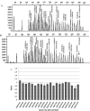
Upper: representative chromatograms from MLPA analysis of CCM1 and CCM3 (salsa MLPA P131 CCM probe mix). A, Results for control individual with peaks corresponding to CCM1 and CCM3 exonic probes. All unlabeled peaks represent the control peaks resulting from the amplification of probes located on different chromosomes. B, Chromatogram from CCM affected proband; Arrow marks the peak corresponding to exon 18 of CCM1, which shows a relative reduction in the peak area in the proband compared with the control. Lower: C. A quantitative analysis demonstrates that the relative peak area has decreased to ∼50%. CCM1 exon 18 deletion is represented, in the graph, by smaller bar marked with arrow. In this graph, the probes located on different chromosomes are not shown and the order of CCM1 and CCM3 exonic probes in C (bars) does not coincide with that shown in A and B (peaks).
Figure 4
Figure 4
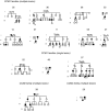
Pedigrees of families with CCM1, CCM2 and CCM3 mutations. Pedigrees are arranged as reported in Table 2; no pedigrees were available for probands 56 and 28. formula image = proband; formula image formula image = affected; formula image formula image = asymptomatic; formula image formula image = obligate carrier; formula image formula image = not known to be affected; M = mutation; WT = no mutation; formula image formula image = deceased.

References

    1. Akers AL, Johnson E, Steinberg GK, Zabramski JM, Marchuk DA (2009) Biallelic somatic and germline mutations in cerebral cavernous malformations (CCMs): evidence for a two‐hit mechanism of CCM pathogenesis. Hum Mol Genet 18:919–930. - PMC - PubMed
    1. Battistini S, Rocchi R, Cerase A (2007) Clinical, magnetic resonance imaging, and genetic study of 5 Italian families with cerebral cavernous malformation. Arch Neurol 64:843–848. - PubMed
    1. Bergametti F, Denier C, Labauge P, Arnoult M, Boetto S, Clanet M et al; Société Française de Neurochirurgie (2005) Mutations within the programmed cell death 10 gene cause cerebral cavernous malformations. Am J Hum Genet 76:42–51. - PMC - PubMed
    1. Cartegni L, Chew SL, Krainer AR (2002) Listening to silence and understanding nonsense: exonic mutations that affect splicing. Nat Rev Genet 3:285–298. - PubMed
    1. Cau M, Loi M, Melis M, Congiu R, Loi A, Meloni C et al (2009) C329X in KRIT1 is a founder mutation among CCM patients in Sardinia. Eur J Med Genet 52:344–348. - PubMed

Publication types