

Artificial skin in perspective: concepts and applications
- PMID: 21029393
- PMCID: PMC3021617
- DOI: 10.1111/j.1755-148X.2010.00786.x
Artificial skin in perspective: concepts and applications
Abstract
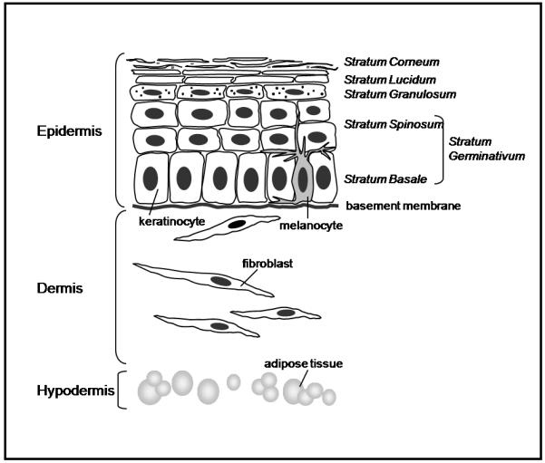
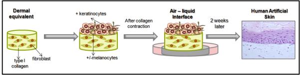
Skin, the largest organ of the human body, is organized into an elaborate layered structure consisting mainly of the outermost epidermis and the underlying dermis. A subcutaneous adipose-storing hypodermis layer and various appendages such as hair follicles, sweat glands, sebaceous glands, nerves, lymphatics, and blood vessels are also present in the skin. These multiple components of the skin ensure survival by carrying out critical functions such as protection, thermoregulation, excretion, absorption, metabolic functions, sensation, evaporation management, and aesthetics. The study of how these biological functions are performed is critical to our understanding of basic skin biology such as regulation of pigmentation and wound repair. Impairment of any of these functions may lead to pathogenic alterations, including skin cancers. Therefore, the development of genetically controlled and well characterized skin models can have important implications, not only for scientists and physicians, but also for manufacturers, consumers, governing regulatory boards and animal welfare organizations. As cells making up human skin tissue grow within an organized three-dimensional (3D) matrix surrounded by neighboring cells, standard monolayer (2D) cell cultures do not recapitulate the physiological architecture of the skin. Several types of human skin recombinants, also called artificial skin, that provide this critical 3D structure have now been reconstructed in vitro. This review contemplates the use of these organotypic skin models in different applications, including substitutes to animal testing.
© 2010 John Wiley & Sons A/S.
Figures
References
-
- Auxenfans C, Fradette J, Lequeux C, Germain L, Kinikoglu B, Bechetoille N, Braye F, Auger FA, Damour O. Evolution of three dimensional skin equivalent models reconstructed in vitro by tissue engineering. Eur J Dermatol. 2008;19:107–13. - PubMed
-
- Balasubramani M, Kumar TR, Babu M. Skin substitutes: a review. Burns. 2001;27:534–44. - PubMed
-
- Barker CL, McHale MT, Gillies AK, Waller J, Pearce DM, Osborne J, Hutchinson PE, Smith GM, Pringle JH. The development and characterization of an in vitro model of psoriasis. J Invest Dermatol. 2004;123:892–901. - PubMed
Publication types
MeSH terms
Grants and funding
LinkOut - more resources
Full Text Sources
Other Literature Sources
Research Materials
