Review
. 2010 Oct 28:7:290.
doi: 10.1186/1743-422X-7-290.

Mobile DNA elements in T4 and related phages

Affiliations
Review

Mobile DNA elements in T4 and related phages

David R Edgell et al. Virol J. .

Abstract

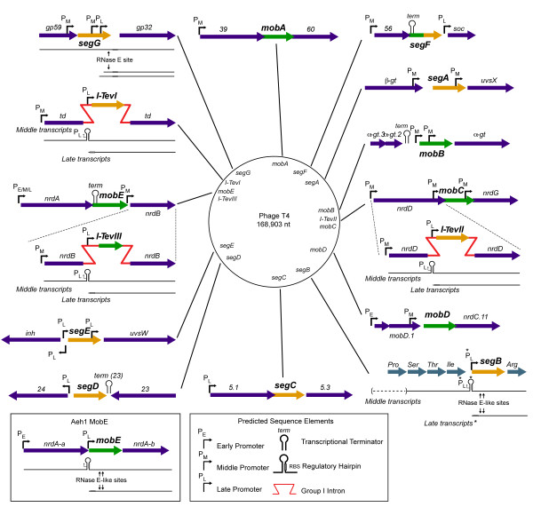
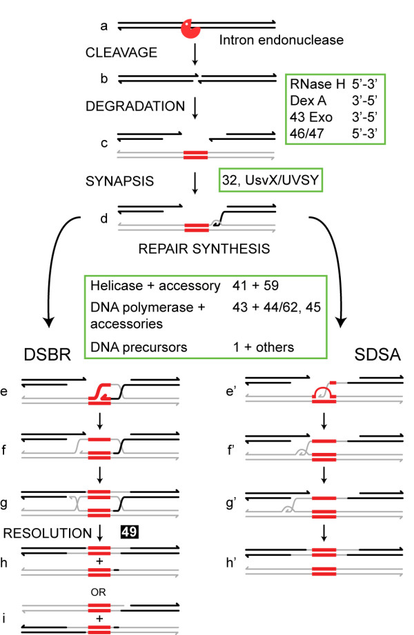
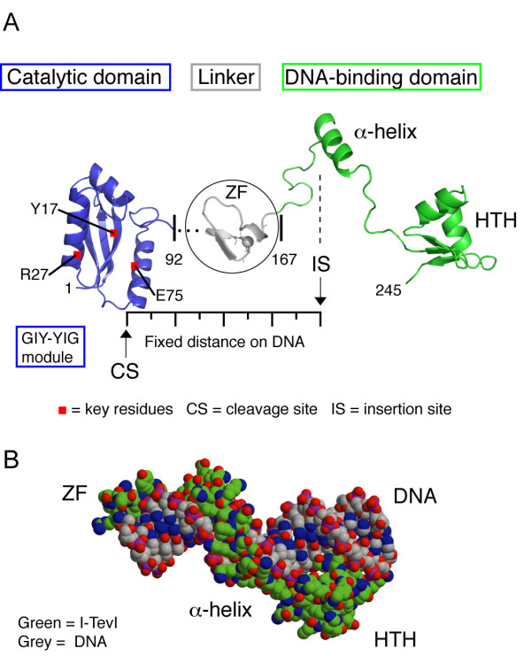
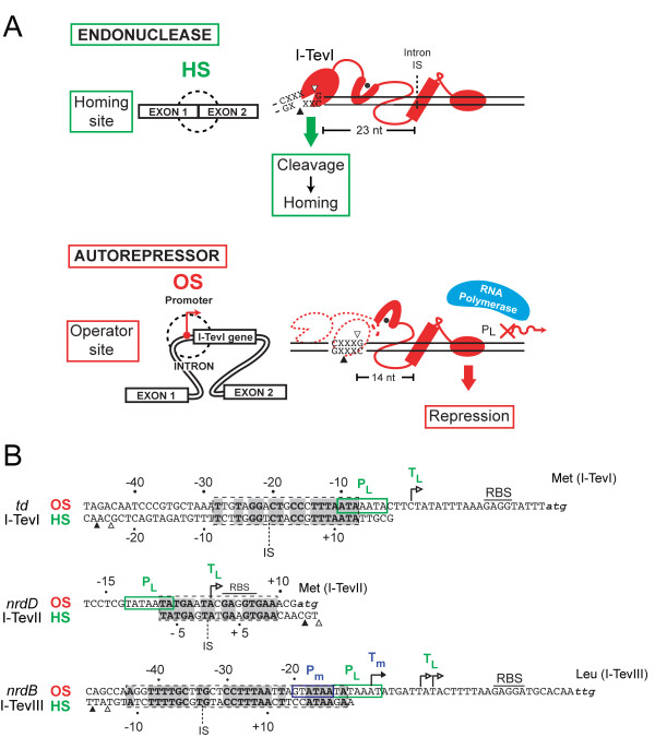
Mobile genetic elements are common inhabitants of virtually every genome where they can exert profound influences on genome structure and function in addition to promoting their own spread within and between genomes. Phage T4 and related phage have long served as a model system for understanding the molecular mechanisms by which a certain class of mobile DNA, homing endonucleases, promote their spread. Homing endonucleases are site-specific DNA endonucleases that initiate mobility by introducing double-strand breaks at defined positions in genomes lacking the endonuclease gene, stimulating repair and recombination pathways that mobilize the endonuclease coding region. In phage T4, homing endonucleases were first discovered as encoded within the self-splicing td, nrdB and nrdD introns of T4. Genomic data has revealed that homing endonucleases are extremely widespread in T-even-like phage, as evidenced by the astounding fact that ~11% of the T4 genome encodes homing endonuclease genes, with most of them located outside of self-splicing introns. Detailed studies of the mobile td intron and its encoded endonuclease, I-TevI, have laid the foundation for genetic, biochemical and structural aspects that regulate the mobility process, and more recently have provided insights into regulation of homing endonuclease function. Here, we summarize the current state of knowledge regarding T4-encoded homing endonucleases, with particular emphasis on the td/I-TevI model system. We also discuss recent progress in the biology of free-standing endonucleases, and present areas of future research for this fascinating class of mobile genetic elements.


Figures

Figure 1
Figure 1
Schematic of the location of the fifteen homing endonuclease genes indicated on a genomic map of bacteriophage T4. For simplicity, each genomic segment is drawn with the endonuclease in the same orientation, with relevant regulatory elements indicated. The GIY-YIG endonucleases are shown in yellow, while the HNH-type endonucleases are green. The hybrid endonuclease segF is drawn with both colours. The bacteriophage Aeh1 mobE endonuclease, which is not part of the bacteriophage T4 genome, is set in a box. An asterix (*) marks a predicted late promoter upstream of SegB.
Figure 2
Figure 2
Alternative mechanisms for DSB-mediated intron homing. Subsequent to cleavage by the homing endonuclease (a), the recipient, intronless allele (thick lines) undergoes exonucleolytic degradation and homologous sequence alignment with an intron-containing donor (thin lines) (b, c). A 3' end of the recipient invades the donor, which serves as a template for repair synthesis (d). In the DSBR pathway (left), DNA synthesis through the intron (red) results in formation and expansion of a D-loop (e), which then serves as substrate for repair synthesis of the noninvading strand (f). Holliday junctions are resolved to produce either noncrossover (h) or crossover (i) products. During synthesis-dependent strand annealing (SDSA) (right), the displaced loop or bubble migrates with the replicative end as DNA synthesis proceeds through the intron (e' - g'). The newly synthesized strand is released from the donor and serves as template for repair synthesis of the noninvading strand (g' - h') to generate noncrossover products only (h'). Functions implicated in homing and their putative association with appropriate steps in the homing pathways are shown.
Figure 3
Figure 3
I-TevI structure. A. Two domains of the enzyme joined by a linker. The catalytic GIY-YIG domain (blue) is separated from the DNA binding domain (green) by a 75-amino acid linker, which includes the zinc finger (grey). The DNA binding domain consists of elongated segments, an α-helix and a helix-turn-helix (HTH) module. B. Space filling model of the DNA-binding domain and zinc finger on DNA. The protein is bound to a 20-bp DNA substrate.
Figure 4
Figure 4
Dual function of I-TevI. A. I-TevI binds with equal affinity to the homing site (top) and operator site (bottom). The CS sequence at the natural distance in the homing site allows endonuclease cleavage, to initiate homing. In the operator site, there is no cleavage sequence at a suitable distance, resulting in autorepression, because I-TevI binding blocks the late promoter and transcription. B. Autorepression by T4 intron-encoded endonucleases. For each of the three endonucleases, I-TevI, I-TevII, and I-TevIII, the endonuclease's homing site (HS) is aligned with proven or putative the operator site (OS) upstream of the endonuclease ORF within the td, nrdB, and nrdD introns, respectively. The operator sites are indicated by dashed boxes, with bold-type nucleotides representing identity between the operator and homing sites. The position of the endonuclease's cleavage sites are indicated by open and black triangles. Green and blue boxes indicate late and middle T4 promoters, respectively, with corresponding transcription start sites indicated by right-facing arrows labeled with the same color.
Figure 5
Figure 5
RNA structures involved in translational regulation of homing endonucleases in T-even bacteriophage. Arrows and red type indicate late promoter position and direction of transcription in the corresponding DNA sequence. For cases where the initating nucleotide has been mapped, it is indicated with a star. The RBS and start codon are shown in blue and black boldface type, respectively. Nucleotide variants in the I-TevI hairpin are indicated for phages TuIa and U5, and the alternative structure of the phage U5 I-TevIII hairpin is indicated by a box. The lower arrows indicate the general genetic organization of hairpin-regulated homing endonucleases. From left to right; the free-standing homing endonucleases mobE and segB, the intron-encoded endonucleases I-TevI, I-TevII, and I-TevIII, and the unusual hairpin for seg43(25) in Aeromonas phage 25.

References

    1. Belfort M. Phage T4 introns: self-splicing and mobility. Annu Rev Genet. 1990;24:363–385. doi: 10.1146/annurev.ge.24.120190.002051. - DOI - PubMed
    1. Huang YJ, Parker MM, Belfort M. Role of exonucleolytic degradation in group I intron homing in phage T4. Genetics. 1999;153:1501–1512. - PMC - PubMed
    1. Mueller JE, Clyman J, Huang YJ, Parker MM, Belfort M. Intron mobility in phage T4 occurs in the context of recombination-dependent DNA replication by way of multiple pathways. Genes Dev. 1996;10:351–364. doi: 10.1101/gad.10.3.351. - DOI - PubMed
    1. Parker MM, Belisle M, Belfort M. Intron homing with limited exon homology. Illegitimate double-strand-break repair in intron acquisition by phage T4. Genetics. 1999;153:1513–1523. - PMC - PubMed
    1. Parker MM, Court DA, Preiter K, Belfort M. Homology requirements for double-strand break-mediated recombination in a phage lambda-td intron model system. Genetics. 1996;143:1057–1068. - PMC - PubMed

Publication types

MeSH terms

LinkOut - more resources