

Distinct neuroanatomical substrates and cognitive mechanisms of figure copy performance in Alzheimer's disease and behavioral variant frontotemporal dementia
- PMID: 21029744
- PMCID: PMC3005024
- DOI: 10.1016/j.neuropsychologia.2010.10.026
Distinct neuroanatomical substrates and cognitive mechanisms of figure copy performance in Alzheimer's disease and behavioral variant frontotemporal dementia
Abstract
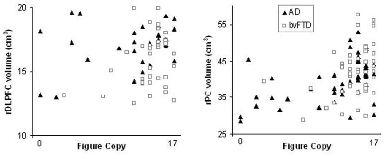
Figure copy is the most common method of visual spatial assessment in dementia evaluations, but performance on this test may be multifactorial. We examined the neuroanatomical substrates of figure copy performance in 46 patients with Alzheimer's disease (AD) and 48 patients with the behavioral variant of Frontotemporal dementia (bvFTD). A group of 94 neurologically healthy controls were studied for comparison. In AD, poor figure copy correlated significantly with right parietal cortex volumes but not with right dorsolateral prefrontal cortex volumes, whereas in bvFTD, figure copy performance correlated significantly with right dorsolateral prefrontal cortex volumes and there was only a trend with right parietal cortex volumes. The cognitive processes associated with figure copy performance also differed by diagnostic group such that figure copy was associated with spatial perception and attention in AD and with spatial planning and working memory in bvFTD. Spatial planning accounted for unique variance in the figure copy performance of bvFTD even after accounting for spatial perception, attention, and working memory. These results suggest that figure copy performance in AD and bvFTD is not anatomically specific and is differentially impacted by bottom-up and top-down aspects of visual spatial processing. Alternative methods of visual spatial assessment for dementia evaluations are proposed.
Copyright © 2010 Elsevier Ltd. All rights reserved.
Figures
References
-
- Barton JJ. Higher cortical visual function. Curr Opin Ophthalmol. 1998;9(6):40–45. - PubMed
-
- Boxer AL, Rankin KP, Miller BL, Schuff N, Weiner M, Gorno-Tempini ML, et al. Cinguloparietal atrophy distinguishes Alzheimer disease from semantic dementia. Arch Neurol. 2003;60(7):949–956. - PubMed
-
- Braak H, Braak E. Evolution of neuronal changes in the course of Alzheimer’s disease. J Neural Transm Suppl. 1998;53:127–140. - PubMed
-
- Buckner RL, Head D, Parker J, Fotenos AF, Marcus D, Morris JC, et al. A unified approach for morphometric and functional data analysis in young, old, and demented adults using automated atlas-based head size normalization: reliability and validation against manual measurement of total intracranial volume. Neuroimage. 2004;23(2):724–738. - PubMed
Publication types
MeSH terms
Grants and funding
LinkOut - more resources
Full Text Sources
Medical
