. 2011 Apr;1812(4):544-8.
doi: 10.1016/j.bbadis.2010.10.010. Epub 2010 Oct 26.

Iron loading-induced aggregation and reduction of iron incorporation in heteropolymeric ferritin containing a mutant light chain that causes neurodegeneration

Affiliations

Iron loading-induced aggregation and reduction of iron incorporation in heteropolymeric ferritin containing a mutant light chain that causes neurodegeneration

Barry B Muhoberac et al. Biochim Biophys Acta. 2011 Apr.

Abstract

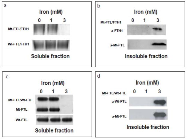
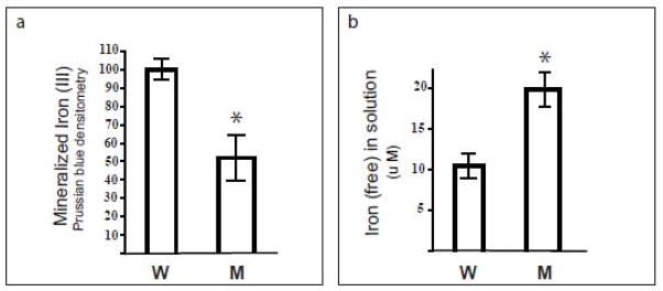
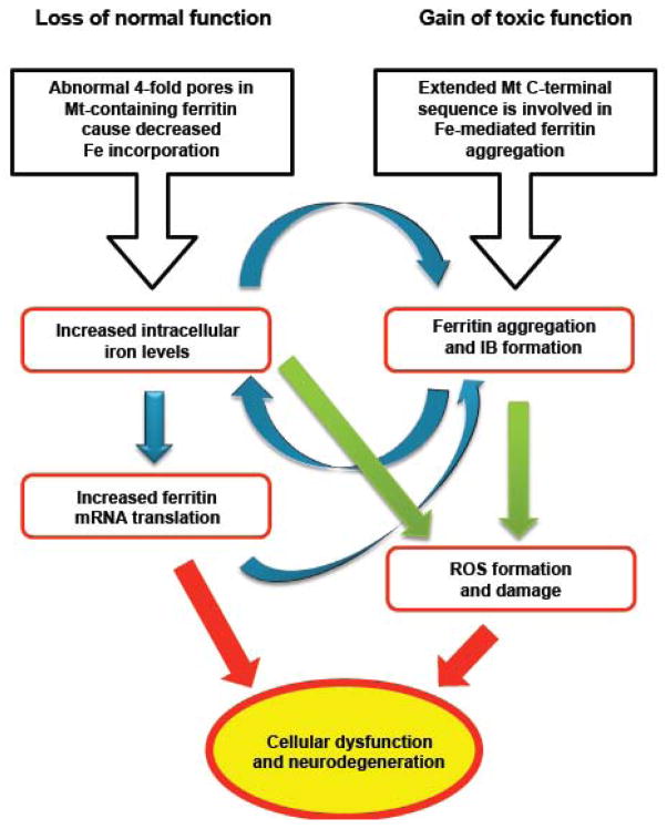
Hereditary ferritinopathy (HF) is a neurodegenerative disease characterized by intracellular ferritin inclusion bodies (IBs) and iron accumulation throughout the central nervous system. Ferritin IBs are composed of mutant ferritin light chain as well as wild-type light (Wt-FTL) and heavy chain (FTH1) polypeptides. In vitro studies have shown that the mutant light chain polypeptide p.Phe167SerfsX26 (Mt-FTL) forms soluble ferritin 24-mer homopolymers having a specific structural disruption that explains its functional problems of reduced ability to incorporate iron and aggregation during iron loading. However, because ferritins are usually 24-mer heteropolymers and all three polypeptides are found in IBs, we investigated the properties of Mt-FTL/FTH1 and Mt-FTL/Wt-FTL heteropolymeric ferritins. We show here the facile assembly of Mt-FTL and FTH1 subunits into soluble ferritin heteropolymers, but their ability to incorporate iron was significantly reduced relative to Wt-FTL/FTH1 heteropolymers. In addition, Mt-FTL/FTH1 heteropolymers formed aggregates during iron loading, contrasting Wt-FTL/FTH1 heteropolymers and similar to what was seen for Mt-FTL homopolymers. The resulting precipitate contained both Mt-FTL and FTH1 polypeptides as do ferritin IBs in patients with HF. The presence of Mt-FTL subunits in Mt-FTL/Wt-FTL heteropolymers also caused iron loading-induced aggregation relative to Wt-FTL homopolymers, with the precipitate containing Mt- and Wt-FTL polypeptides again paralleling HF. Our data demonstrate that co-assembly with wild-type subunits does not circumvent the functional problems caused by mutant subunits. Furthermore, the functional problems characterized here in heteropolymers that contain mutant subunits parallel those problems previously reported in homopolymers composed exclusively of mutant subunits, which strongly suggests that the structural disruption characterized previously in Mt-FTL homopolymers occurs in a similar manner and to a significant extent in both Mt-FTL/FTH1 and Mt-FTL/Wt-FTL heteropolymers.


Figures

Figure 1
Figure 1. Ferritin structural disruption and aggregation caused by Mt-FTL subunits
(a) The structure of the spherical shell is maintained in mutant ferritin as seen in thecrystallographic structures of Wt- and Mt-FTL homopolymers viewed down one of their 4-fold axes. (b) Close up views of the 4-fold pore from the interior of the Wt- and Mt-FTL structures show remarkable disruption in Mt-FTL making the pores unstable and leaky. Note that since the last 26 amino acids of Mt-FTL remained unaccounted for crystallographically, mutant C-termini are substantially longer than represented in (b), and if extended could reach as far as the diameter of the ferritin shell. (c) Iron loading-induced aggregation of Mt-FTL homopolymers is consistent with a model in which iron binds to the unraveled and extended portion of the mutant C-termini on two different ferritin shells bridging them and initiating a gradual accumulation of ferritin and iron into a precipitate. Bridging is not necessarily restricted to C-termini and may become more general, e.g., between a C-terminal group and a surface amino acid which both have affinity for iron. Structures were taken from RCSB (code 2FG8 for Wt-FTL and 3HX2 for Mt-FTL) and the precipitation model was modified from [14].
Figure 2
Figure 2. Formation of ferritin heteropolymers from Mt-FTL and FTH1 polypeptides
Recombinant Mt-FTL and FTH1 (or Wt-FTL and FTH1) polypeptides (1 mM) were mixed in a 1:1 ratio and allowed to co-assemble into heteropolymers by gradually removing GdnHCl. (a) Five g of reassembled Mt-FTL/FTH1 or Wt-FTL/FTH1 apoferritin heteropolymers were resolved by 3–8% native PAGE and stained with Coomasie blue. Mt-FTL, FTH1, and Wt-FTL apoferritin homopolymers were run as controls. (b) Size exclusion chromatographic elution profiles of the heteropolymers were obtained with a Superose 6 10/300 GL column with elution time markers for molecular weights indicated with arrows.
Figure 3
Figure 3. Iron loading-induced precipitation of ferritin heteropolymers containing Mt-FTL subunits
Ferrous ammonium sulphate was added to separate samples of heteropolymeric (Mt-FTL/FTH1, Wt-FTL/FTH1, and Mt-FTL/Wt-FTL) or homopolymeric (Mt-FTL and Wt-FTL) apoferritins, and incubated for 2 h. The protein concentration was 1 μM and iron concentrations were 0, 1, and 3 mM. Samples were centrifuged for 15 min at 10,000 g in order to separate soluble from aggregated protein. In contrast to the solubility of Wt-FTL/FTH1 heteropolymers, precipitation of Mt-FTL/FTH1 and Mt-FTL/Wt-FTL heteropolymers was observed at 3 mM iron. (a) Soluble fractions of Mt-FTL/FTH1 and Wt-FTL/FTH1 heteropolymers were characterized by 3–8% native PAGE and stained with Coomasie blue. (b) The insoluble fraction was resuspended in Laemmli sample buffer, resolved by SDS 12% PAGE, and blotted with antibodies against FTH1 and Mt-FTL. (c) Mt-FTL/Wt-FTL heteropolymers and the corresponding homopolymers were iron loaded and electrophoresed as in (a) above, showing precipitation for hetero- or homopolymers containing Mt-FTL. (d) The insoluble fraction from Mt-FTL/Wt-FTL was resuspended, electrophoresed, and blotted as in (b) above, with antibodies against Wt-FTL and Mt-FTL.
Figure 4
Figure 4. Iron incorporation by soluble ferritin heteropolymers
Mt-FTL/FTH1 (M) and Wt-FTL/FTH1 (W) apoferritin heteropolymers were separately incubated with ferrous ammonium sulphate at concentrations of 1 μM and 1 mM, respectively. After 2 h, incubation was stopped by the addition of HCl and bathophenanthroline. (a) After separating unincorporated iron from the protein by electrophoresis on non-denaturing gels (3–8% native PAGE), iron biomineral within the heteropolymer interior was quantitated as density of Prussian blue formed in the protein bands. (b) Iron incorporation by the Mt-FTL/FTH1 heteropolymers relative to Wt-FTL/FTH1 was determined through measurement of residual ferrous iron in solution. Bathophenantholine was added to the samples after the 2 h incubation with iron, the samples were diluted 10-fold with buffer, their absorbances were read at 535 nm, and the amount of chelated Fe(II) was calculated using an extinction coefficient of 22140 M−1cm −1.
Figure 5
Figure 5. Altered iron metabolism and ferritin aggregation in the pathogenesis of HF
Iron homeostasis is crucial in providing an optimum amount of iron as a cofactor for proteins while preventing generation of ROS, which can cause damage to proteins, membranes and DNA [1]. The cellular abnormalities associated with the FTL mutation originate in 4-fold pore disruption and enhanced aggregation, and can be understood in terms of a loss of normal function (left) and a gain of toxic function (right) of ferritin. Increased cellular iron levels, up-regulation of ferritin production, and aggregation appear to work synergistically (blue arrows) to form large nuclear and cytoplasmic IBs, perhaps even causing cellular misfunction through mechanical crowding. Increased iron levels may enhance ROS generation at a location separate from ferritin or as part of the compromised iron handling and aggregation of ferritin itself (green arrows). Thus cellular dysfunction may be a consequence of both a decrease in normal function and an increase of toxic function of ferritin leading to neurodegeneration.

References

    1. Zecca L, Youdim MB, Riederer P, Connor JR, Crichton RR. Iron, brain ageing and neurodegenerative disorders. Nat Rev Neurosci. 2004;5:863–873. - PubMed
    1. Crichton R. Iron metabolism from molecular mechanisms to clinical consequences. 3. Wiley; West Sussex, U.K: 2009.
    1. Stefanini S, Cavallo S, Wang CQ, Tataseo P, Vecchini P, Giartosio A, Chiancone E. Thermal stability of horse spleen apoferritin and human recombinant H apoferritin. Arch Biochem Biophys. 1996;325:58–4. - PubMed
    1. Santambrogio P, Levi S, Arosio P, Palagi L, Vecchio G, Lawson DM, Yewdall SJ, Artymiuk PJ, Harrison PM, Jappelli R, Cesareni G. Evidence that a salt bridge in the light chain contributes to the physical stability difference between heavy and light human ferritins. J Biol Chem. 1992;267:14077–14083. - PubMed
    1. Levi S, Santambrogio P, Cozzi A, Rovida E, Corsi B, Tamborini E, Spada S, Albertini A, Arosio P. The role of the L-chain in ferritin iron incorporation. Studies of homo and heteropolymers. J Mol Biol. 1994;238:649–654. - PubMed

Publication types