. 2011 Mar;173(3):530-40.
doi: 10.1016/j.jsb.2010.10.014. Epub 2010 Oct 26.

The beginning of a beautiful friendship: cross-linking/mass spectrometry and modelling of proteins and multi-protein complexes

Affiliations

The beginning of a beautiful friendship: cross-linking/mass spectrometry and modelling of proteins and multi-protein complexes

Juri Rappsilber. J Struct Biol. 2011 Mar.

Abstract

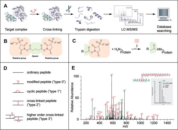
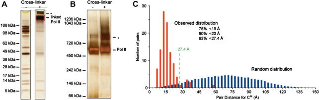
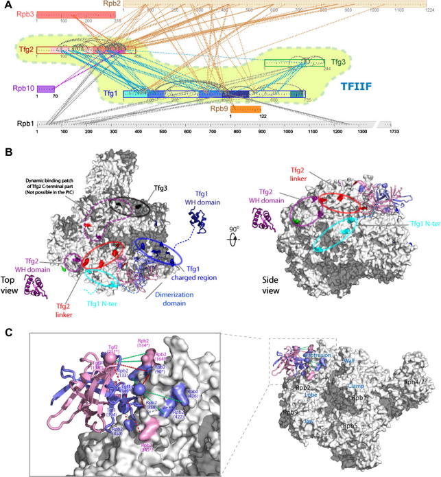
After more than a decade of method development, cross-linking in combination with mass spectrometry and bioinformatics is finally coming of age. This technology now provides improved opportunities for modelling by mapping structural details of functional complexes in solution. The structure of proteins or protein complexes is ascertained by identifying amino acid pairs that are positioned in close proximity to each other. The validity of this technique has recently been benchmarked for large multi-protein complexes, by comparing cross-link data with that from a crystal structure of RNA polymerase II. Here, the specific nature of this cross-linking data will be discussed to assess the technical challenges and opportunities for model building. We believe that once remaining technological challenges of cross-linking/mass spectrometry have been addressed and cross-linking/mass spectrometry data has been incorporated into modelling algorithms it will quickly become an indispensable companion of protein and protein complex modelling and a corner-stone of integrated structural biology.


Figures

Fig.1
Fig.1
(A) Outline of the cross-linking/mass spectrometry process. A target complex is cross-linked in solution and digested with trypsin into peptides. The peptides are analysed by liquid chromatography coupled high-resolution mass spectrometry (LC–MS/MS) to obtain high-resolution masses and fragment masses (high/high) for cross-linked peptides. The fragmentation spectra of all peptides are subjected to database searching to identify cross-linked peptides. As an optional step, cross-linked peptides can be enriched before their LC–MS analysis. (B) A typical cross-linker, here bis(sulfosuccinimidyl)glutarate (BS2G), is composed of two reactive groups on either end separated by a spacer. This cross-linker reacts with primary amines (lysine side chain, protein N-terminus). Others target thiols (cysteine side chain) or activate carboxylic acids (aspartate, glutamate, protein C-terminus) for reaction with primary amines. (C) Reaction of a cross-linker with a primary amine. Part of the cross-linker, the leaving group, is replaced by the primary amine to form a covalent bond between the spacer and the amine. In this case, a peptide bond is formed. R can stand for either the rest of the cross-linker or may contain another protein, if the cross-linker had already reacted on its other end. (D) Peptides types that can be observed after cross-linking and trypsin digestion. (E) High resolution fragmentation spectrum of a cross-linked peptide obtained on an LTQ-Orbitrap mass spectrometer (adapted from (Chen et al., 2010)). Fragment peaks are annotated in red or green, depending on the peptide that fragmented and following the naming convention for peptides (y, C-terminal fragment; b, N-terminal fragment; both as a result of dissociating the peptide bond in the peptide back bone, followed by the number of amino acids included in the fragment and the charge of the fragment). All observed fragments are also indicated as bond cleavages between amino acids in the two cross-linked peptides. In this case, virtually all possible fragments of the peptide pair have been matched and virtually all peaks have been annotated resulting in a high-confidence identification of this cross-link. The inset shows a zoom onto one fragment peak (m/z 576, 3248) which matched with −1.1 ppm to the proposed peptide sequence. The high resolution of the spectrum (R 7505 for this peak) allows clear separation of the isotope peaks and consequently assignment of the fragment’s charge state.
Fig.2
Fig.2
Benchmarking the cross-linking/mass spectrometry process using S. cerevisiae RNA polymerase II (Pol II) and its crystal structure. (A) The subunits of Pol II are separated by denaturating gel electrophoresis (SDS–PAGE) and visualized by silver staining. The individual subunits can be seen as separate bands before the addition of cross-linker (here bis(sulfosuccinimidyl)suberate (BS3)). After cross-linking, these individual bands disappear and a new, high-molecular weight band appears, corresponding to the cross-linked Pol II (red box). A higher molecular weight band corresponds possibly to Pol II dimers (asterisk). (B) Pol II migrates under native conditions mostly as a single band, both in the absence and presence of cross-linking. Under both conditions, some Pol II dimerization is observed (asterisk). (C) Distribution of alpha-carbon distances for lysine pairs in the crystal structure of Pol II (PDB 1WCM) (Armache et al., 2005) when scaling the distance distribution for all random lysine pairs in the crystal structure to 106 pairs (blue) and when taking the distance measure of those 106 pairs that were observed by cross-linking (red) (Chen et al., 2010). The predicted upper limit for cross-linkable lysine pairs in the crystal structure is here 27.4 Å. This upper limit includes the length of lysine side chains (2 × 6.5 Å), the length of the spacer (max. 11.4 Å) and an estimation of the positional error in the crystal structure (1.5 Å for surface residues). The upper limit does not consider the possibility of conformation changes or vibrations of the complex in solution. The observed distribution of cross-linked pairs is clearly not random and fulfils largely the theoretically predicted distance threshold for cross-linkable pairs. (All adapted from (Chen et al., 2010).)
Fig.3
Fig.3
Cross-linking/mass spectrometry analysis of S. cerevisiae RNA polymerase II (Pol II) bound to transcription factor IIF (TFIIF). (A) Linkage map showing the sequence position of all observed cross-linked residue pairs within TFIIF and between TFIIF and Pol II. Connections between residues are blue within TFIIF or colour coded by Pol II subunit for cross-links between Pol II and TFIIF. Sequence regions of TFIIF subunits are colour coded (Tfg1: N-terminal tail, 2 × dimerization domain, charged region, winged-helix (WH) domain; Tfg2: 2 × dimerization domain, linker, WH domain). (B) Residues of Pol II colour coded by region in TFIIF subunits they cross-link with. (C) Homology model of the Tfg1–Tfg2 dimerization domain positioned on the Pol II structure (PDB 1WCM) with cross-linked residues labelled by proteins and residue number. Dashed lines connect pairs of residues that were used for the positioning, either because they were observed to cross-link or because they are the closest residues in the structure (denoted by an asterisk behind their residue number). (All adapted from (Chen et al., 2010).)

References

    1. Aebersold R., Mann M. Mass spectrometry-based proteomics. Nature. 2003;422:198–207. - PubMed
    1. Alber F., Dokudovskaya S., Veenhoff L.M., Zhang W., Kipper J., Devos D., Suprapto A., Karni-Schmidt O., Williams R., Chait B.T., Rout M.P., Sali A. Determining the architectures of macromolecular assemblies. Nature. 2007;450:683–694. - PubMed
    1. Alber F., Dokudovskaya S., Veenhoff L.M., Zhang W., Kipper J., Devos D., Suprapto A., Karni-Schmidt O., Williams R., Chait B.T., Sali A., Rout M.P. The molecular architecture of the nuclear pore complex. Nature. 2007;450:695–701. - PubMed
    1. Armache K.J., Mitterweger S., Meinhart A., Cramer P. Structures of complete RNA polymerase II and its subcomplex, Rpb4/7. J. Biol. Chem. 2005;280:7131–7134. - PubMed
    1. Back J.W., de Jong L., Muijsers A.O., de Koster C.G. Chemical cross-linking and mass spectrometry for protein structural modeling. J. Mol. Biol. 2003;331:303–313. - PubMed

Publication types

LinkOut - more resources