. 2010 Oct 29;143(3):390-403.
doi: 10.1016/j.cell.2010.09.049.

The long noncoding RNA, Jpx, is a molecular switch for X chromosome inactivation

Affiliations

The long noncoding RNA, Jpx, is a molecular switch for X chromosome inactivation

Di Tian et al. Cell. .

Abstract

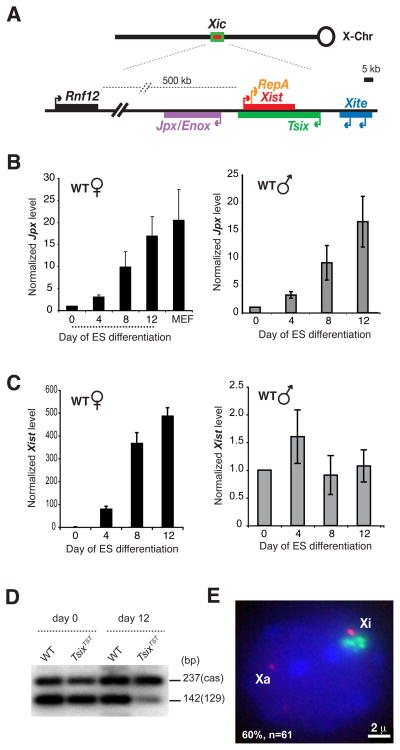
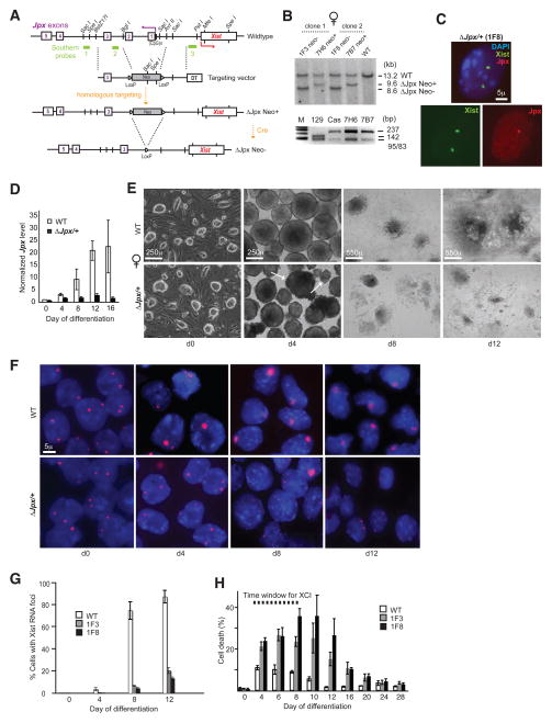
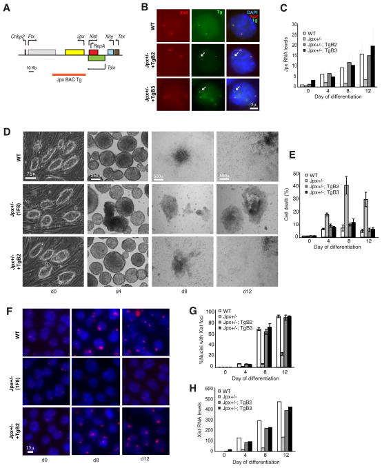
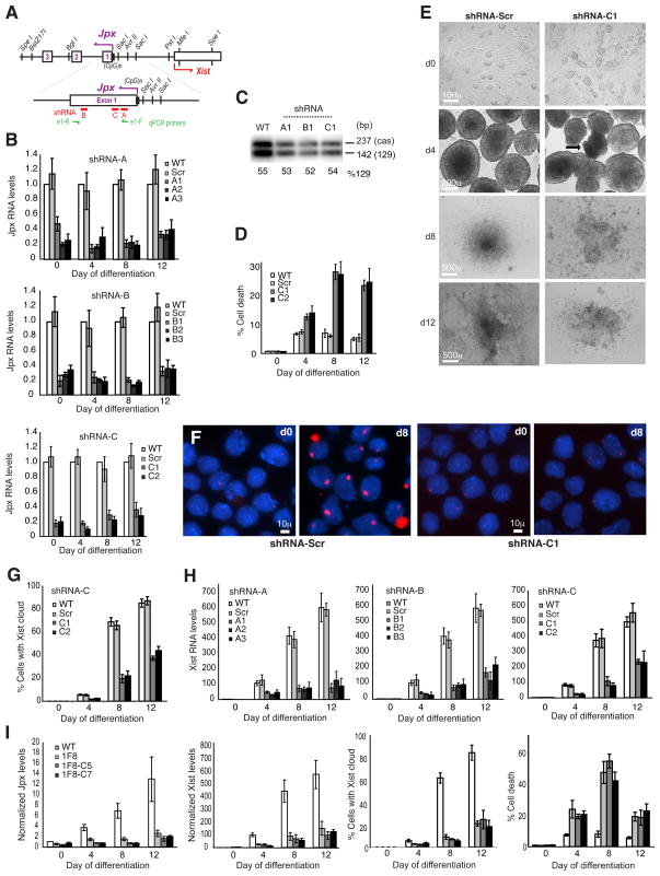
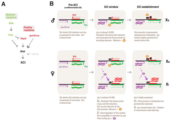
Once protein-coding, the X-inactivation center (Xic) is now dominated by large noncoding RNAs (ncRNA). X chromosome inactivation (XCI) equalizes gene expression between mammalian males and females by inactivating one X in female cells. XCI requires Xist, an ncRNA that coats the X and recruits Polycomb proteins. How Xist is controlled remains unclear but likely involves negative and positive regulators. For the active X, the antisense Tsix RNA is an established Xist repressor. For the inactive X, here, we identify Xic-encoded Jpx as an Xist activator. Jpx is developmentally regulated and accumulates during XCI. Deleting Jpx blocks XCI and is female lethal. Posttranscriptional Jpx knockdown recapitulates the knockout, and supplying Jpx in trans rescues lethality. Thus, Jpx is trans-acting and functions as ncRNA. Furthermore, ΔJpx is rescued by truncating Tsix, indicating an antagonistic relationship between the ncRNAs. We conclude that Xist is controlled by two RNA-based switches: Tsix for Xa and Jpx for Xi.


Figures

Figure 1
Figure 1. Jpx expression increases 10- to 20-fold during ES cell differentiation
(A) The Xic and its noncoding genes. Rnf12 is coding and lies 500 kb away. (B) Time-course analyses of Jpx expression by qRT-PCR in differentiating female and male ES cells. Averages and standard error (S.E.) from three (female) or four (male) independent differentiation experiments are plotted. Values are normalized to Gapdh RNA and d0 Jpx levels are set to 1.0. (C) Time-course analyses of Xist expression by qRT-PCR in differentiating male and female ES cells. Averages and S.E. from 6 (male) and 3 (female) independent differentiation experiments are plotted. All values are normalized to Gadph RNA and d0 Xist is set to 1.0. (D) Allele-specific RT-PCR analysis of Jpx in wildtype and TsixTST/+ female ES cells on d0 and d12 of differentiation (E) RNA FISH indicates that Jpx escapes inactivation in 60% of d16 female cells. N=61. Xist clouds are present in 98% of cells. Xist RNA, green. Jpx RNA, red.
Figure 2
Figure 2. ΔJpx causes loss of XCI and massive cell death in female ES cells
(A) The Jpx gene, targeting vector, and products of homologous targeting before and after Cre-mediated excision of the Neo positive-selection marker. DT, diphtheria toxin for negative selection. (CpG)n, CpG island. Numbered boxes represent five Jpx exons. (B) Top panel: Southern analysis of SacI-digested genomic DNA from ΔJpx/+ and WT female ES cells using probe 1. The Neo- female clones, 1F3 and 1F8, were derived from the Neo+ 6H7 and 7B7 clones, respectively. Bottom panel: Allele-specific PCR analysis showed that the 129 allele was preferentially targeted over the M. castaneus (cas) allele. The analysis for Neo+ 6H7 and 7B7 clones are shown. M, 100-bp markers. (C) DNA FISH of ΔJpx/+ female ES cells. Xist probe (pSx9), FITC-labeled. The Jpx probe (Cy3-labelled, red) is located in the region of deletion. (D) Time-course analyses of Jpx expression by qRT-PCR in differentiating WT and ΔJpx/+ female ES cells. Averages and standard errors (S.E.) from three independent differentiation experiments are plotted, with values normalized first to Gapdh and then d0 WT Jpx levels are set to 1.0. (E) Brightfield photographs of WT and ΔJpx/+ female ES cells from d0 to d12 of differentiation. Arrows point to disintegrating, necrotic EBs present in mutant cultures. (F) RNA FISH to examine the time course of Xist upregulation. Xist probe, Cy3-labelled pSx9. (G) Plotted time course of Xist upregulation in WT and two ΔJpx/+ mutants, 1F3 and 1F8. Averages +/−S.E. from 3 independent differentiation experiments are shown. Sample sizes (n): d0, 595–621; d4, 922–1163; d8, 3013–4370; d12, 3272–4794. (H) Massive cell death in mutant female cells. The Trypan blue staining results of 3 independent differentiation experiments were averaged and plotted with S.E. d0, n=150–800 cells for d0; d4, n=200–500 cells; all other timepoints, n=500–2000 cells.
Figure 3
Figure 3. Transgenic Jpx rescues ΔJpx in trans
(A) Map of the Xic and 90-kb Jpx transgene. (B) Multiprobe DNA FISH to localize Xist (pSx9, red) and Jpx (BAC, green) in two independent transgenic clones, TgB2 and TgB3. Arrows, Jpx transgene. (C) Time-course analyses of Jpx expression by qRT-PCR in differentiating cells of indicated genotype. Averages +/−S.E. from three independent differentiation experiments are plotted. Values are normalized to Gapdh RNA and WT d0 Jpx level is set to 1.0. D) Brightfield photographs of WT and transgenic EB from d0 to d12. (E) Cell death analysis of WT, knockout, and transgenic EB, performed as above. (F) RNA FISH to examine the time course of Xist upregulation. Xist probe, Cy3-labelled pSx9. (G) Quantitation of WT, knockout, and transgenic EB with Xist RNA foci (RNA FISH) from d0–d12. (H) qRT-PCR of steady state Xist levels in WT, knockout, and transgenic EB from d0–d12.
Figure 4
Figure 4. Jpx functions as a long ncRNA
(A) A map of the 5′ end of Jpx showing its exons (purple), shRNA locations, and qPCR primer positions. (B) Significant knockdown of Jpx RNA in 2–3 independent clones for each Jpx-specific shRNA, but not in the scrambled shRNA clone (Scr). Jpx RNA levels are normalized to WT levels for each day of differentiation. A1–A3 are clones for shRNA-A; B1–B3 for shRNA-B; and C1, C2 for shRNA-C. (C) Residual Jpx RNA was extracted from d8 shRNA clones, A1, B1, and C1, and subjected to allele-specific RT-PCR (Nla-III polymorphism). The gel was blotted and hybridized to an end-labelled oligo. Allelic fractionation shows similar ratios of 129:castaneus bands in WT and knockdown clones, suggesting that the shRNAs affected both Jpx alleles. Only 10–30% of Jpx RNA was left in the knockdowns and therefore the PCR was overcycled to visualize the low residual levels of Jpx in the knockdown cells. (D) Cell death assay shows that loss of Jpx RNA reduces cell viability during differentiation. Clones shRNA-C1 and –C2 are shown, but shRNA-A and –B clones also show increased cell death. (E) Brightfield images show poor EB formation and outgrowth in knockdowns but not Scr control. (F) Xist RNA FISH shows loss of Xist upregulation when Jpx is knocked down using shRNA-C. (G) Quantitation of the number of cells with Xist RNA clusters from three independent differentiation experiments of control and knockdown clones. Average +/− S.E. shown. (H) Quantitation of Xist RNA levels in control and knockdown clones from 3 independent differentiation experiments. RNA levels are normalized to d0 WT values. Average +/− S.E. shown. Differentiation of shRNA-A and –B knockdown clones were performed at the same time; therefore, WT and Scr values for shRNA-A and shRNA-B are the same. (I) Jpx knockdown in ΔJpx/+ cells (1F8) using shRNA-C. Independent clones, C5 and C7, behaved similarly to each other and also to their parent, 1F8, in all assays shown. Average +/− S.E. shown. All values are normalized to d0 WT.
Figure 5
Figure 5. Jpx’s mild cis-preference revealed in ΔJpx/+ survivors
(A) Xist RNA FISH on d28 EB. Xist probe, Cy3-labelled pSx9. WT, 97% Xist+ cells (n>2000). ΔJpx/+, 92% Xist+ cells (n>3000). (B) Allele-specific RNA/DNA FISH determines which X is Xi. FITC-labelled pSx9 probe detects Xist RNA and the Xist locus from both Xs, whereas the Cy3-labelled Jpx probe detects only the wildtype X (the probe resides in the deleted region). (C) Percent of 1F8 mutant female cells where Xi = XΔ (i.e., X129). Averages +/−S.E. from 3 independent differentiation experiments. (D) Allele-specific RT-PCR of indicated transcripts from d0–d28. The percentage of transcripts from the 129 allele (%129) is determined by phosphorimaging. +, WT. Δ, 1F8 mutant. Values for lanes that are not visible are obtained after a longer exposure. (E) Two-color RNA FISH for Xist and Pgk1 transcripts in d28 cells. (F) Summary of ΔJpx effects on male and female ES cells.
Figure 6
Figure 6. A Tsix RNA truncation suppresses ΔJpx
(A) Targeting the Tsix truncation mutation (TsixTST) (Ogawa et al., 2008) to the ΔJpx chromosome in 1F8 female ES cells. TsixTST prematurely terminates Tsix at the targeted triple polyA site (trpA) 1 kb downstream of the major Tsix promoter. 1F8-S1 and 1F8-S2 are two independently generated double mutant clones. IRES, internal ribosome entry site. Puro, puromycin selection marker. (B) Southern analysis using EcoRV digestion to confirm targeting. The X129 and Xcas alleles have a ~300 bp DXPas34 length polymorphism. The X129 allele was targeted in both 1F8-S1 and 1F8-S2. (C) Cell death analysis shows that TsixTST partially rescues viability of ΔJpx/+ ES cells. (D) Brightfield photographs of wildtype, single, and double mutant female ES cells during differentiation. (E) RNA FISH indicating that Xist upregulation (large red clouds) is rescued in double mutants. (F) TsixTST restores Xist induction in ΔJpx/+ cells. Averages +/− S.D. shown for three independent differentiation experiments. (G) The pattern of allelic skewing is reversed in ΔJpx; TsixTST/+ cells. (H, I) Further depletion of Jpx RNA by shRNA-C knockdown in ΔJpx; TsixTST/+ cells did not alter the phenotype of the double mutant, as shown by qRT-PCR of Xist expression (H) and by EB outgrowth to d8 (I). Jpx; TsixTST/+, 1F8-S2. Two shRNA-C clones derived from 1F8-S2 were examined (C1, C2).
Figure 7
Figure 7. Model and summary
(A) Proposed epistasis model: Xist is under positive-negative regulation by noncoding genes. Xite and Tsix repress Xist, whereas Jpx and RepA activate Xist. Arrows, positive relationship. Blunt arrows, negative relationship. Rnf12 is a coding gene. (B) Proposed events in male and female ES cells. Xist silencers (orange hexagons) include Dnmt3a and other chromatin modifications. Jpx (purple oval) is depicted as a diffusible transacting RNA. Open lollipops, unmethylated Xist promoter. Filled lollipops, methylated Xist promoter.

References

    1. Ahn JY, Lee JT. Retinoic acid accelerates downregulation of the Xist repressor, Oct4, and increases the likelihood of Xist activation when Tsix is deficient. BMC Dev Biol. 2010;10:90. - PMC - PubMed
    1. Ariel M, Robinson E, McCarrey JR, Cedar H. Gamete-specific methylation correlates with imprinting of the murine Xist gene. Nat Genet. 1995;9:312–315. - PubMed
    1. Bacher CP, Guggiari M, Brors B, Augui S, Clerc P, Avner P, Eils R, Heard E. Transient colocalization of X-inactivation centres accompanies the initiation of X inactivation. Nat Cell Biol. 2006;8:293–299. - PubMed
    1. Brockdorff N, Ashworth A, Kay GF, McCabe VM, Norris DP, Cooper PJ, Swift S, Rastan S. The product of the mouse Xist gene is a 15 kb inactive X-specific transcript containing no conserved ORF and located in the nucleus. Cell. 1992;71:515–526. - PubMed
    1. Brown CJ, Hendrich BD, Rupert JL, Lafreniere RG, Xing Y, Lawrence J, Willard HF. The human XIST gene: analysis of a 17 kb inactive X-specific RNA that contains conserved repeats and is highly localized within the nucleus. Cell. 1992;71:527–542. - PubMed

Publication types