. 2010 Oct 29;143(3):430-41.
doi: 10.1016/j.cell.2010.09.024.

Endophilin functions as a membrane-bending molecule and is delivered to endocytic zones by exocytosis

Affiliations

Endophilin functions as a membrane-bending molecule and is delivered to endocytic zones by exocytosis

Jihong Bai et al. Cell. .

Abstract

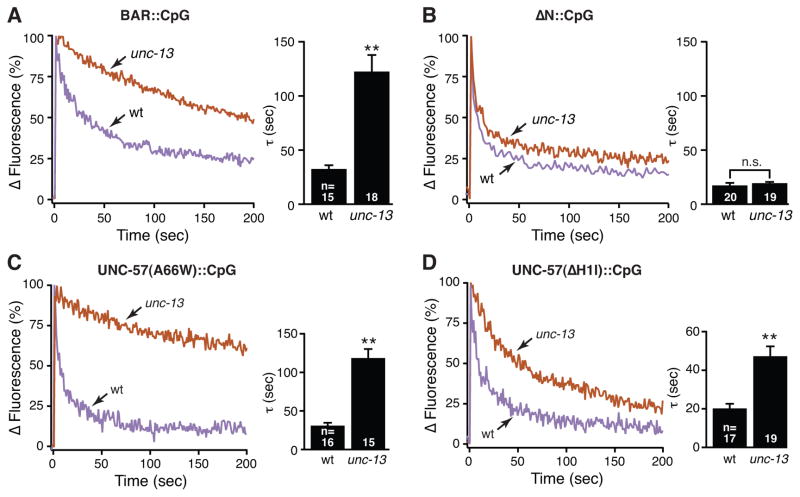
Two models have been proposed for endophilin function in synaptic vesicle (SV) endocytosis. The scaffolding model proposes that endophilin's SH3 domain recruits essential endocytic proteins, whereas the membrane-bending model proposes that the BAR domain induces positively curved membranes. We show that mutations disrupting the scaffolding function do not impair endocytosis, whereas those disrupting membrane bending cause significant defects. By anchoring endophilin to the plasma membrane, we show that endophilin acts prior to scission to promote endocytosis. Despite acting at the plasma membrane, the majority of endophilin is targeted to the SV pool. Photoactivation studies suggest that the soluble pool of endophilin at synapses is provided by unbinding from the adjacent SV pool and that the unbinding rate is regulated by exocytosis. Thus, endophilin participates in an association-dissociation cycle with SVs that parallels the cycle of exo- and endocytosis. This endophilin cycle may provide a mechanism for functionally coupling endocytosis and exocytosis.


Figures

Figure 1
Figure 1. The UNC-57 BAR domain promotes SV endocytosis through its membrane interactions
[See also Figure S1 and S2] The phenotypes of wild type (wt), unc-57(e406) endophilin mutants, and the indicated transgenic strains were compared. Transgenes were mCherry tagged UNC-57 variants including full length (FL; residues 1-379), BAR domain (residues 1-283) and N (residues 27-379). Transgenes were expressed in all neurons, using the snb-1 promoter. Expression levels of these transgenes are shown in Figure S1. (A) Representative one minute locomotion trajectories are shown (n= 20 animals for each genotype). The starting points for each trajectory were aligned for clarity. (B) Locomotion rates are compared for the indicated genotypes. Representative traces (C) and summary data for endogenous EPSC rates (D) are shown. Representative images (E) and summary data (F) for axonal SpH fluorescence in the dorsal nerve cord are shown for the indicated genotypes. The number of worms analyzed for each genotype is indicated. (**) indicates p <0.001 compared to WT controls. (##) indicates p< 0.001 when compared to unc-57 mutants. Error bars = SEM.
Figure 2
Figure 2. The membrane-bending activity of Endophilin A BAR domains promotes SV endocytosis
[See also Figure S3] Transgenes encoding wild type and mutant BAR domains (1–247) from rat EndophilinA1 (rEndoA1 BAR) were analyzed for their ability to rescue locomotion rate (A), the surface Synaptobrevin (SpH) (Figure S3B) and EPSC rate (BC) defects of unc-57 mutants. The ΔH1, A66W, and M70S/I71S mutations alter membrane tubulation activity but have little or no effect on membrane binding in vitro (Gallop et al., 2006; Masuda et al., 2006). All transgenes were tagged with mCherry at the C-terminus to assess differences in expression levels (Figure S3). (D) Transgenes expressing BAR domains derived from different proteins were compared for their ability to rescue the locomotion rate defect of unc-57 mutants. BAR domains are indicated as follows: rat Endophilin A (rEndo A1, A2, and A3; residues 1-247), lamprey Endophilin A (LampEndo; residues 1-248), C. elegans (CeEndo B; residues 1-267), rat Endophilin B (rEndo B; residues 1-247), and rat Amphiphysin (rAmphiphysin; residues 1-250). (E) Alignment of the H1 helix sequence is shown for the indicated BAR domains. The A66 residue (green, arrow) is required for tubulation activity (Masuda et al., 2006). rEndo A3 has a sequence polymorphism (S64Y) compared to the A1 and A2 isoforms. (F) Rescuing activities of rEndo A1, A2, A3, and A3(Y64S) BAR domains for the unc-57 mutant locomotion defect are compared. All transgenes were expressed in all neurons using the snb-1 promoter. The number of animals analyzed for each genotype is indicated. (**) and (*) indicate significant differences compared to wt (p< 0.001 and p< 0.01, respectively). (##) indicates p< 0.001 when compared to unc-57 mutants. Error bars = SEM.
Figure 3
Figure 3. Endophilin is targeted to the SV pool at presynaptic terminals
[See also Figure S4] Full-length unc-57 Endophilin was tagged at the C-terminus with mCherry and photoactivatable GFP (designated as CpG) (schematic shown in Fig. 4A). (AB) The distribution of UNC-57::CpG mcherry fluorescence in DA neuron dorsal axons is compared with a co-expressed SV (GFP::SNB-1, A) or endocytic marker (APT-4::GFP AP2α, B). (C) Targeting of UNC-57::CpG to presynaptic terminals was strongly reduced in unc-104(e1265) KIF1A mutants.
Figure 4
Figure 4. Exocytosis promotes dissociation of Endophilin from the SV pool
[See also Figure S5] (A). Photoactivation of UNC-57::CpG at a single synapse is shown schematically (above) and in representative images (below). (BC). Representative images and traces of photoactivated UNC-57::CpG green fluorescence decay in wild type (wt) and unc-13(s69) mutants. The mCherry fluorescence was captured to control for motion artifacts. (D). Dispersion rates of photoactivated UNC-57::CpG were quantified in the indicated genotypes. Decay constants (τ) are 28.1 ± 3.3 sec for wt; 117.2 ± 13.6 sec for unc-13 (s69); 136.2 ± 26.4 sec for unc-18 (e81); and 68.2 ± 6.5 sec for tom-1(nu468)unc-13(s69). Representative images (E) and summary data (F) for steady-state UNC-57::CpG mCherry fluorescence in the dorsal nerve cord axons was compared for the indicated genotypes. (F) Synaptic enrichment of UNC-57::CpG was calculated as follows: ΔF/F = (FpeakFaxon)/Faxon. The number of animals analyzed for each genotype is indicated. ** indicates p <0.001 compared to wt controls. Error bars = SEM.
Figure 5
Figure 5. Structural requirements for UNC-57 regulation by exocytosis
[See also Figure S6] Representative traces and summary data are shown comparing the dispersion of mutant UNC-57 proteins. Mutant proteins analyzed are: (A) WT BAR domain lacking the SH3 (BAR reporter), and full length UNC-57 proteins containing the ΔN (membrane binding deficient) (B), A66W (tubulation deficient) (C), and ΔH1I (dimerization deficient) (D) mutations. Each mutant protein was tagged with CpG, expressed in DA neurons, and their dispersion rates compared following photoactivation in wild type and unc-13 mutants. (**) indicates p <0.001 compared to wt controls. Error bars = SEM.
Figure 6
Figure 6. RAB-3 and the Rab3 GEF (AEX-3) regulate Endophilin targeting to SVs
Representative images (A) and quantification (B) of UNC-57::CpG synaptic enrichment in wt, aex-3, unc-13 and unc-13;aex-3 double mutants were shown (Synaptic enrichment: wt 8.6 ± 0.6; aex-3 6.2 ± 0.6; unc-13; aex-3 19.6 ± 1.2, unc-13 32.1 ± 2.2 fold). Dispersion rates of UNC-57::CpG in wt (τ = 28.1 ± 3.3 sec) and aex-3 mutant (τ = 45.5 ± 5.1 sec) animals were compared in (C). (DE) UNC-57::CpG distribution in transgenic unc-13 mutant animals with over-expressed RAB-3 (Q81L) or (T36N) was studied. Over-expression of RAB-3 (Q81L), but not RAB-3 (T36N) significantly reduced UNC-57::CpG synaptic enrichment in unc-13 mutants. ** indicates p <0.001 and * indicates p <0.01, compared to wt controls. Error bars = SEM.
Figure 7
Figure 7. Analysis of a membrane-anchored UNC-57 protein
[See also Figure S7] (A) The distribution of UNC-57(PM) in DA neuron axons was compared with co-expressed UNC-57::CpG (upper panels), active zone [AZ] marker ELKS-1::mcherry (middle panels), or endocytic zone [EZ] marker APT-4::mcherry (AP2α, lower panels). UNC-57(PM) comprises full length UNC-57 and GFP fused to the N-terminus of UNC-64 Syntaxin 1A (schematic shown in Figure S7A). (B) GFP fluorescence of UNC-57(PM) in wt and unc-13(s69) mutant animals were quantified. UNC-57(PM) was expressed in all neurons with the snb-1 promoter. (C) Representative images are shown of wild type and mutant UNC-57(PM) proteins in dorsal cord axons. The BAR(PM) protein corresponds to UNC-57(PM) lacking the SH3 domain. The ΔN(PM) protein lacks the N-terminal 26 residues of UNC-57 (which prevents membrane binding). (D) Representative traces of endogenous EPSC from wt, unc-57(e406) mutants, and transgenic unc-57 animals carrying wild type and mutant UNC-57(PM) constructs. Endogenous EPSC rates (left panel) and amplitudes (right panel) are shown in (E). Significant differences (p <0.001 by student’s t-test) are indicated as: **, compared to wt; and ##, compared to unc-57 mutants. Error bars = SEM.

References

    1. Ch’ng Q, Sieburth D, Kaplan JM. Profiling synaptic proteins identifies regulators of insulin secretion and lifespan. PLoS Genet. 2008;4:e1000283. - PMC - PubMed
    1. Darcy KJ, Staras K, Collinson LM, Goda Y. Constitutive sharing of recycling synaptic vesicles between presynaptic boutons. Nat Neurosci. 2006;9:315–321. - PubMed
    1. Dickman DK, Horne JA, Meinertzhagen IA, Schwarz TL. A slowed classical pathway rather than kiss-and-run mediates endocytosis at synapses lacking synaptojanin and endophilin. Cell. 2005;123:521–533. - PubMed
    1. Dittman J, Ryan TA. Molecular circuitry of endocytosis at nerve terminals. Annu Rev Cell Dev Biol. 2009;25:133–160. - PubMed
    1. Dittman JS, Kaplan JM. Factors regulating the abundance and localization of synaptobrevin in the plasma membrane. Proc Natl Acad Sci U S A. 2006;103:11399–11404. - PMC - PubMed

Publication types

Substances