

doi: 10.1242/dmm.002915.
Towards improved animal models of neonatal white matter injury associated with cerebral palsy
Affiliations
- PMID: 21030421
- PMCID: PMC2965396
- DOI: 10.1242/dmm.002915
Item in Clipboard
Towards improved animal models of neonatal white matter injury associated with cerebral palsy
Dis Model Mech.
2010 Nov-Dec.
Abstract
Newborn neurological injuries are the leading cause of intellectual and motor disabilities that are associated with cerebral palsy. Cerebral white matter injury is a common feature in hypoxic-ischemic encephalopathy (HIE), which affects full-term infants, and in periventricular leukomalacia (PVL), which affects preterm infants. This article discusses recent efforts to model neonatal white matter injury using mammalian systems. We emphasize that a comprehensive understanding of oligodendrocyte development and physiology is crucial for obtaining new insights into the pathobiology of HIE and PVL as well as for the generation of more sophisticated and faithful animal models.
Figures
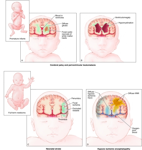
Common types of injury associated with development of CP in ELBW and term infants. (A,B) Illustration of brain injuries commonly affecting ELBW infants. (A) Key characteristics of intraventricular hemorrhage, which results from germinal matrix hemorrhage into the ventricles, sometimes extending into the brain parenchyma, are shown. Additionally, there is a high incidence of PVL (a type of WMI), which can result in a cystic necrosis of white matter tracts and/or diffuse gliosis. (B) Long-term sequelae of brain injury in ELBW infants are shown, including hypomyelination resulting from failure of lesion repair, as well as ventriculomegaly, which represents an ex-vacuo change resulting from significant loss of brain parenchyma. (C,D) Common brain injuries in full-term infants. (C) Neonatal stroke in which a focal region of cortex is affected. (D) HIE results in global HI injury to the brain, more specifically to neurons of the cortical plate and basal ganglia, as well as white matter tracts.
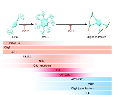
Markers of oligodendrocyte lineage specification and maturation. The schematic shows oligodendrocyte lineage progression from OPCs to preOLs and then to myelinating oligodendrocytes. In the past decade, various markers have been identified that show lineage- and stage-specific expression (indicated by colored gradients). The markers PDGFRα, Olig2, Nkx2.2, Sox10, NG2 and Olig1 (nuclear) (indicated in orange) are characteristic of OPCs. O4 and O1 [also known as galactocebroside (GalC)] (indicated in red) mark intermediate preOLs, whereas APC (also known as CC1), myelin basic protein (MBP), myelin proteolipid protein (PLP) and Olig1 (cytoplasmic) (indicated in blue) are typical of mature myelinating oligodendrocytes. Several laboratories studying MS and PVL have reported abnormal oligodendrocyte differentiation in these disorders, suggesting that these abnormalities are associated with a failure to repair demyelinated lesions.
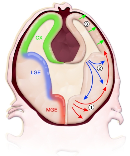
Oligodendrocytes are generated in multiple waves and in various locations of the CNS. Results from the Richardson laboratory and others have shown that, in mice, oligodendrocytes of the forebrain are initially specified in ventral regions, such as the medial ganglionic eminence (MGE; corresponding to region 1 in the figure), a ventral domain of embryonic proliferative precursor cells, from which the oligodendrocytes migrate to more dorsal areas of the brain (Kessaris et al., 2006). At subsequent developmental stages, later waves of oligodendrocytes are produced in successively more dorsal regions, such as the lateral ganglionic eminence (LGE, corresponding to region 2) in late embryonic development and the cerebral cortex (CX, corresponding to region 3) in the early postnatal period. The first three waves of oligodendrocyte production are shown. In addition, NG2-positive precursor cells continue to cycle in the adult brain and are thought to contribute to oligodendrocyte turnover, as well as the response to injury (not shown). It is unclear whether oligodendrocytes from different regions of the brain have cell-intrinsic differences in their ability to repair myelin, or in their vulnerability to HI or other toxic insults.
Similar articles
-
The vulnerable oligodendrocyte: inflammatory observations on a cause of cerebral palsy.Neurology. 2001 May 22;56(10):1254-5. doi: 10.1212/wnl.56.10.1254. Neurology. 2001. PMID: 11376167 Review. No abstract available.
-
Diffusion tensor imaging in children with periventricular leukomalacia: variability of injuries to white matter tracts.AJNR Am J Neuroradiol. 2007 Aug;28(7):1213-22. doi: 10.3174/ajnr.A0534. AJNR Am J Neuroradiol. 2007. PMID: 17698519 Free PMC article.
-
Nitrosative and oxidative injury to premyelinating oligodendrocytes in periventricular leukomalacia.J Neuropathol Exp Neurol. 2003 May;62(5):441-50. doi: 10.1093/jnen/62.5.441. J Neuropathol Exp Neurol. 2003. PMID: 12769184
-
Cross-sectional comparison of periventricular leukomalacia in preterm and term children.Neurology. 2010 Apr 27;74(17):1386-91. doi: 10.1212/WNL.0b013e3181dad62d. Neurology. 2010. PMID: 20421583
-
Periventricular leukomalacia: overview and recent findings.Pediatr Dev Pathol. 2006 Jan-Feb;9(1):3-13. doi: 10.2350/06-01-0024.1. Epub 2006 Apr 4. Pediatr Dev Pathol. 2006. PMID: 16808630 Review.
Cited by
-
Impact of early brain lesions on the optic radiations in children with cerebral palsy.Front Neurosci. 2022 Oct 5;16:924938. doi: 10.3389/fnins.2022.924938. eCollection 2022. Front Neurosci. 2022. PMID: 36278011 Free PMC article.
-
Behavioral and histological outcomes following neonatal HI injury in a preterm (P3) and term (P7) rodent model.Behav Brain Res. 2014 Feb 1;259:85-96. doi: 10.1016/j.bbr.2013.10.038. Epub 2013 Nov 1. Behav Brain Res. 2014. PMID: 24185032 Free PMC article.
-
White matter injury in the preterm infant: pathology and mechanisms.Acta Neuropathol. 2017 Sep;134(3):331-349. doi: 10.1007/s00401-017-1718-6. Epub 2017 May 22. Acta Neuropathol. 2017. PMID: 28534077 Free PMC article. Review.
-
Axin2 as regulatory and therapeutic target in newborn brain injury and remyelination.Nat Neurosci. 2011 Jun 26;14(8):1009-16. doi: 10.1038/nn.2855. Nat Neurosci. 2011. PMID: 21706018 Free PMC article.
-
Early-stage effect of HIBD on neuro-motor function and organic composition of neurovascular units in neonatal rats.Front Neurosci. 2023 Nov 21;17:1242936. doi: 10.3389/fnins.2023.1242936. eCollection 2023. Front Neurosci. 2023. PMID: 38075277 Free PMC article.
References
-
- Alix JJ, Fern R. (2009). Glutamate receptor-mediated ischemic injury of premyelinated central axons. Ann Neurol. 66, 682–693 - PubMed
-
- Althaus HH, Kloppner S, Klopfleisch S, Schmitz M. (2008). Oligodendroglial cells and neurotrophins: a polyphonic cantata in major and minor. J Mol Neurosci. 35, 65–79 - PubMed
-
- Anjari M, Counsell SJ, Srinivasan L, Allsop JM, Hajnal JV, Rutherford MA, Edwards AD. (2009). The association of lung disease with cerebral white matter abnormalities in preterm infants. Pediatrics 124, 268–276 - PubMed
-
- Anthony TE, Heintz N. (2007). The folate metabolic enzyme ALDH1L1 is restricted to the midline of the early CNS, suggesting a role in human neural tube defects. J Comp Neurol. 500, 368–383 - PubMed
-
- Arnett HA, Fancy SP, Alberta JA, Zhao C, Plant SR, Kaing S, Raine CS, Rowitch DH, Franklin RJ, Stiles CD. (2004). bHLH transcription factor Olig1 is required to repair demyelinated lesions in the CNS. Science 306, 2111–2115 - PubMed
Publication types
MeSH terms
Grants and funding
LinkOut - more resources
Full Text Sources
Other Literature Sources
Medical
