. 2011 Jan 20;117(3):915-9.
doi: 10.1182/blood-2010-08-303305. Epub 2010 Oct 28.

Discovery of recurrent t(6;7)(p25.3;q32.3) translocations in ALK-negative anaplastic large cell lymphomas by massively parallel genomic sequencing

Affiliations

Discovery of recurrent t(6;7)(p25.3;q32.3) translocations in ALK-negative anaplastic large cell lymphomas by massively parallel genomic sequencing

Andrew L Feldman et al. Blood. .

Abstract

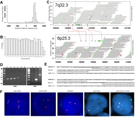
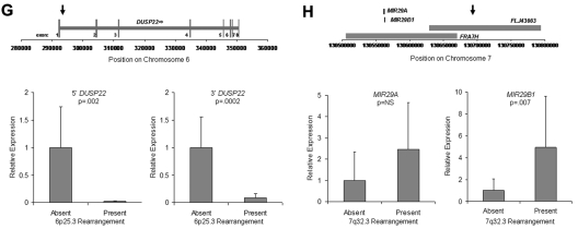
The genetics of peripheral T-cell lymphomas are poorly understood. The most well-characterized abnormalities are translocations involving ALK, occurring in approximately half of anaplastic large cell lymphomas (ALCLs). To gain insight into the genetics of ALCLs lacking ALK translocations, we combined mate-pair DNA library construction, massively parallel ("Next Generation") sequencing, and a novel bioinformatic algorithm. We identified a balanced translocation disrupting the DUSP22 phosphatase gene on 6p25.3 and adjoining the FRA7H fragile site on 7q32.3 in a systemic ALK-negative ALCL. Using fluorescence in situ hybridization, we demonstrated that the t(6;7)(p25.3;q32.3) was recurrent in ALK-negative ALCLs. Furthermore, t(6;7)(p25.3;q32.3) was associated with down-regulation of DUSP22 and up-regulation of MIR29 microRNAs on 7q32.3. These findings represent the first recurrent translocation reported in ALK-negative ALCL and highlight the utility of massively parallel genomic sequencing to discover novel translocations in lymphoma and other cancers.


Figures

Figure 1
Figure 1
Discovery of recurrent t(6;7)(p25.3;q32.3) translocations in ALK-negative ALCLs using mate-pair library sequencing. (A) Histogram demonstrating the calculated distances between the sequenced ends of mate-pairs in which both ends mapped to the reference genome. Most mate-pairs map approximately 5000 bp apart. (B) Bridged coverage of the genome by chromosome, where “bridged” coverage represents the portion of the genome analyzed for the presence of translocations and includes both the sequenced mate-pairs and the intervening DNA segments (∼ 5000 bp total per mate-pair). Average bridged coverage for the genome was approximately 7x. (C) Schematic representation of mapping of mate-pairs to the reference genome. The region of the known 6p25.3 rearrangement is shown in the bottom panel. The x-axis shows nucleotides according to the February 2009 genome assembly (GRCh37/hg19). Horizontal black bars represent approximately 5000-bp DNA fragments between sequenced mate-pairs, which are colored green and red to represent positive and negative strands, respectively. Blue dots represent mate-pairs in which the 2 ends map to the same chromosome more than 25 000 bp apart. Colored numerals represent mate-pairs in which the 2 ends map to different chromosomes, with the color indicating strand, the number indicating the partner chromosome, and the position along the right-hand y-axis representing the position along the partner chromosome. Ten distinct (ie, nonidentical) mate-pairs map to both 6p25.3 and a narrow region of chromosome 7: 8 (green) on the positive strand and 2 (red) on the negative strand. (Top panel) The corresponding locus on 7q32.3. Because these aberrant mate-pairs have ends that map to distinct regions of the genome, they suggest a possible translocation between loci adjacent to these paired ends. Occasional single numerals (eg, the red “14” in the lower right of the bottom panel) represent sporadic, nonrepeated, aberrant mate-pairs that may be introduced during the ligation step of the mate-pair library preparation. The putative breakpoints lie between the positive-strand mate-pairs (green) and the negative-strand mate-pairs (red). Vertical lines indicate the actual breakpoints confirmed by PCR and sequencing. (D) Gel electrophoreses of PCR reactions amplifying the DNA regions containing the putative breakpoints on der(6) and der(7). Each set of reactions included a single chromosome 6 primer and multiple chromosome 7 primers (supplemental Table 1) at increasing predicted distances from the putative breakpoint. (E) Bands shown in panel D were conventionally sequenced and aligned to the genome. Breakpoints of der(6) and der(7) are shown with their alignment to the positive strand of 6p25.3 (DUSP22) and the negative strand of 7q32.3. Nineteen bases of der(7) do not align but reside within a 33-bp sequence (capitalized) complementary to the positive strand of DUSP22 at the 6p25.3 breakpoint (underlined), suggesting a microinversion associated with the breakpoint on 6p25.3. (F) FISH results in the sequenced case (100×/1.40 NA oil objective). The breakapart (BAP) probe to 6p25.3 shows one normal fusion signal and separation of the red and green components of the other signal (arrows), indicating a translocation. This separation of red and green components also is seen with the DUSP22 BAP probe, confirming the 6p25.3 breakpoint involves DUSP22. The additional red signal results from a cross-hybridization to chromosome 16p11. No abnormal separation is seen with the IRF4 BAP probe; again, a cross-hybridization (red signal) is seen. A 7q32.3 BAP probe confirms the presence of a translocation; this signal pattern was seen in 45% of ALK-negative ALCLs with 6p25.3 rearrangements. A dual-fusion (D-) FISH probe showed one normal red signal, one normal green signal, and 2 abnormal fusion signals (arrows), confirming a balanced translocation between 6p25.3 and 7q32.3. (G) The 6p25.3 breakpoint (arrow; top panel) lies within intron 1 of DUSP22. (Bottom panels) DUSP22 expression in ALCLs without and with 6p25.3 rearrangements (real-time quantitative PCR, shown as expression relative to the mean value of the nontranslocated cases, mean ± SD: 5′, 1.00 ± 0.74 vs 0.02 ± 0.01; 3′, 1.00 ± 0.55 vs 0.09 ± 0.08). (H) The 7q32.3 breakpoint (arrow; top panel) lies in the noncoding transcript region FLJ43663, immediately telomeric to the fragile site, FRA7H, and the microRNAs, MIR29A and MIR29B1. (Bottom panels) MicroRNA expression in ALCLs without and with7q32.3 rearrangements (real-time quantitative PCR, shown as expression relative to the mean value of the nontranslocated cases, mean ± SD: MIR29A, 1.00 ± 1.34 vs 2.44 ± 2.20; MIR29B1, 1.00 ± 1.05 vs 4.93 ± 4.68).
Figure 1
Figure 1
Discovery of recurrent t(6;7)(p25.3;q32.3) translocations in ALK-negative ALCLs using mate-pair library sequencing. (A) Histogram demonstrating the calculated distances between the sequenced ends of mate-pairs in which both ends mapped to the reference genome. Most mate-pairs map approximately 5000 bp apart. (B) Bridged coverage of the genome by chromosome, where “bridged” coverage represents the portion of the genome analyzed for the presence of translocations and includes both the sequenced mate-pairs and the intervening DNA segments (∼ 5000 bp total per mate-pair). Average bridged coverage for the genome was approximately 7x. (C) Schematic representation of mapping of mate-pairs to the reference genome. The region of the known 6p25.3 rearrangement is shown in the bottom panel. The x-axis shows nucleotides according to the February 2009 genome assembly (GRCh37/hg19). Horizontal black bars represent approximately 5000-bp DNA fragments between sequenced mate-pairs, which are colored green and red to represent positive and negative strands, respectively. Blue dots represent mate-pairs in which the 2 ends map to the same chromosome more than 25 000 bp apart. Colored numerals represent mate-pairs in which the 2 ends map to different chromosomes, with the color indicating strand, the number indicating the partner chromosome, and the position along the right-hand y-axis representing the position along the partner chromosome. Ten distinct (ie, nonidentical) mate-pairs map to both 6p25.3 and a narrow region of chromosome 7: 8 (green) on the positive strand and 2 (red) on the negative strand. (Top panel) The corresponding locus on 7q32.3. Because these aberrant mate-pairs have ends that map to distinct regions of the genome, they suggest a possible translocation between loci adjacent to these paired ends. Occasional single numerals (eg, the red “14” in the lower right of the bottom panel) represent sporadic, nonrepeated, aberrant mate-pairs that may be introduced during the ligation step of the mate-pair library preparation. The putative breakpoints lie between the positive-strand mate-pairs (green) and the negative-strand mate-pairs (red). Vertical lines indicate the actual breakpoints confirmed by PCR and sequencing. (D) Gel electrophoreses of PCR reactions amplifying the DNA regions containing the putative breakpoints on der(6) and der(7). Each set of reactions included a single chromosome 6 primer and multiple chromosome 7 primers (supplemental Table 1) at increasing predicted distances from the putative breakpoint. (E) Bands shown in panel D were conventionally sequenced and aligned to the genome. Breakpoints of der(6) and der(7) are shown with their alignment to the positive strand of 6p25.3 (DUSP22) and the negative strand of 7q32.3. Nineteen bases of der(7) do not align but reside within a 33-bp sequence (capitalized) complementary to the positive strand of DUSP22 at the 6p25.3 breakpoint (underlined), suggesting a microinversion associated with the breakpoint on 6p25.3. (F) FISH results in the sequenced case (100×/1.40 NA oil objective). The breakapart (BAP) probe to 6p25.3 shows one normal fusion signal and separation of the red and green components of the other signal (arrows), indicating a translocation. This separation of red and green components also is seen with the DUSP22 BAP probe, confirming the 6p25.3 breakpoint involves DUSP22. The additional red signal results from a cross-hybridization to chromosome 16p11. No abnormal separation is seen with the IRF4 BAP probe; again, a cross-hybridization (red signal) is seen. A 7q32.3 BAP probe confirms the presence of a translocation; this signal pattern was seen in 45% of ALK-negative ALCLs with 6p25.3 rearrangements. A dual-fusion (D-) FISH probe showed one normal red signal, one normal green signal, and 2 abnormal fusion signals (arrows), confirming a balanced translocation between 6p25.3 and 7q32.3. (G) The 6p25.3 breakpoint (arrow; top panel) lies within intron 1 of DUSP22. (Bottom panels) DUSP22 expression in ALCLs without and with 6p25.3 rearrangements (real-time quantitative PCR, shown as expression relative to the mean value of the nontranslocated cases, mean ± SD: 5′, 1.00 ± 0.74 vs 0.02 ± 0.01; 3′, 1.00 ± 0.55 vs 0.09 ± 0.08). (H) The 7q32.3 breakpoint (arrow; top panel) lies in the noncoding transcript region FLJ43663, immediately telomeric to the fragile site, FRA7H, and the microRNAs, MIR29A and MIR29B1. (Bottom panels) MicroRNA expression in ALCLs without and with7q32.3 rearrangements (real-time quantitative PCR, shown as expression relative to the mean value of the nontranslocated cases, mean ± SD: MIR29A, 1.00 ± 1.34 vs 2.44 ± 2.20; MIR29B1, 1.00 ± 1.05 vs 4.93 ± 4.68).

References

    1. Rowley JD. Chromosomal translocations: revisited yet again. Blood. 2008;112(6):2183–2189. - PubMed
    1. Swerdlow S, Campo E, Harris N, et al., editors. WHO Classification of Tumours of Haematopoietic and Lymphoid Tissues. 4th ed. Lyon, France: International Agency for Research on Cancer; 2008.
    1. Gascoyne RD, Aoun P, Wu D, et al. Prognostic significance of anaplastic lymphoma kinase (ALK) protein expression in adults with anaplastic large cell lymphoma. Blood. 1999;93(11):3913–3921. - PubMed
    1. Li R, Morris SW. Development of anaplastic lymphoma kinase (ALK) small-molecule inhibitors for cancer therapy. Med Res Rev. 2008;28(3):372–412. - PubMed
    1. Mason DY, Harris NL, Delsol G, et al. Anaplastic large cell lymphoma, ALK-negative. In: Swerdlow S, Campo E, Harris N, et al., editors. WHO Classification of Tumours of Haematopoietic and Lymphoid Tissues. 4th ed. Lyon, France: International Agency for Research on Cancer; 2008. pp. 317–319.

Publication types

MeSH terms

Substances