. 2011 Jan 7;286(1):370-9.
doi: 10.1074/jbc.M110.179499. Epub 2010 Oct 28.

Liver X receptor (LXR) regulates human adipocyte lipolysis

Affiliations

Liver X receptor (LXR) regulates human adipocyte lipolysis

Britta M Stenson et al. J Biol Chem. .

Abstract

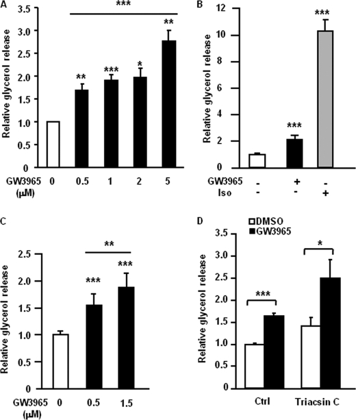
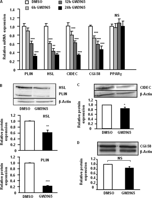
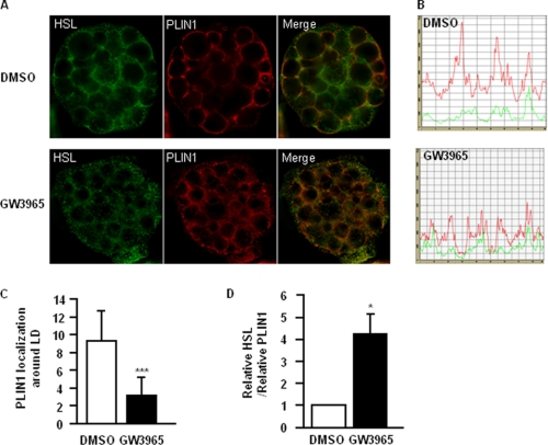
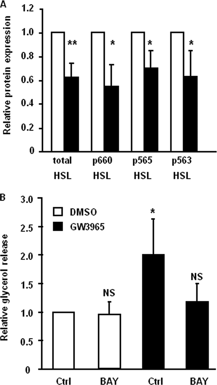
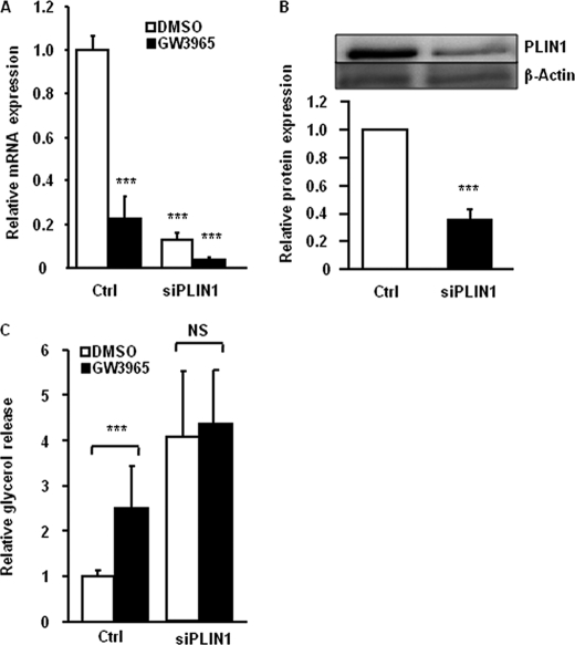
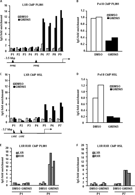
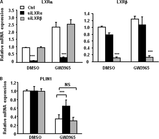
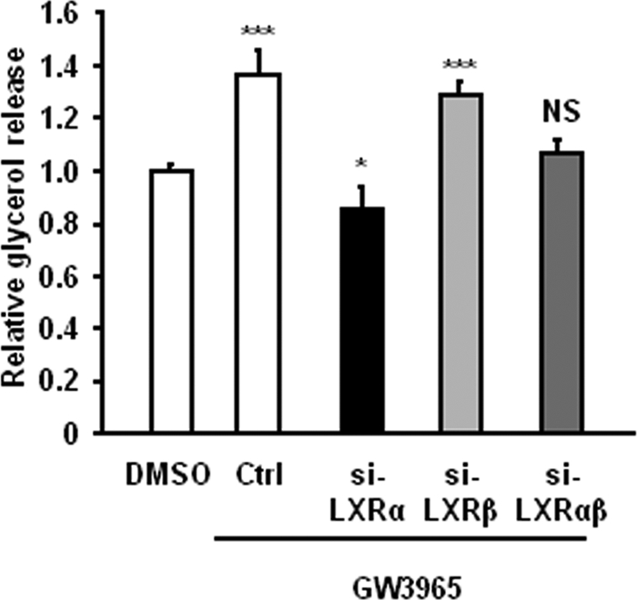
The Liver X receptor (LXR) is an important regulator of carbohydrate and lipid metabolism in humans and mice. We have recently shown that activation of LXR regulates cellular fuel utilization in adipocytes. In contrast, the role of LXR in human adipocyte lipolysis, the major function of human white fat cells, is not clear. In the present study, we stimulated in vitro differentiated human and murine adipocytes with the LXR agonist GW3965 and observed an increase in basal lipolysis. Microarray analysis of human adipocyte mRNA following LXR activation revealed an altered gene expression of several lipolysis-regulating proteins, which was also confirmed by quantitative real-time PCR. We show that expression and intracellular localization of perilipin1 (PLIN1) and hormone-sensitive lipase (HSL) are affected by GW3965. Although LXR activation does not influence phosphorylation status of HSL, HSL activity is required for the lipolytic effect of GW3965. This effect is abolished by PLIN1 knockdown. In addition, we demonstrate that upon activation, LXR binds to the proximal regions of the PLIN1 and HSL promoters. By selective knock-down of either LXR isoform, we show that LXRα is the major isoform mediating the lipolysis-related effects of LXR. In conclusion, the present study demonstrates that activation of LXRα up-regulates basal human adipocyte lipolysis. This is at least partially mediated through LXR binding to the PLIN1 promoter and down-regulation of PLIN1 expression.


Figures

FIGURE 1.
FIGURE 1.
LXR up-regulates glycerol release in human and murine 3T3-L1 adipocytes independent of re-esterification. A, in vitro-differentiated human adipocytes were treated with different concentrations of GW3965 (black bars) or vehicle (white bar) for 48 h, after which medium was removed, and glycerol release was measured. Values were corrected for protein concentration in each experimental group. Means ± S.D. *, p < 0.05; **, p < 0.01; ***, p < 0.001; n = 3–7. B, adipocyte cultures were pretreated with 1 μm of GW3965 (black bar) or vehicle (DMSO, white bar) for 48 h and incubated in lipolytic medium for 3 h. Glycerol release was measured, and values were corrected for protein concentration in each experimental group. The known lipolytic agent isoprenaline (Iso) was used as a positive control for acute lipolysis induction. Means ± S.D., n = 3. C, differentiated 3T3-L1 adipocytes were treated with different concentrations of GW3965 (black bars) or vehicle (white bar) for 48 h, after which medium was removed, and glycerol release was measured. Values were corrected for protein concentration in each experimental group. Means ± S.D. **, p < 0.01; ***, p < 0.001, n = 7. D, adipocyte cultures were pretreated with 1 μm of GW3965 (black bars) or vehicle (DMSO, white bars) for 48 h, and incubated in lipolytic medium for 3 h with or without Triacsin C. Glycerol release was measured, and values were corrected for protein concentration in each experimental group. Means ± S.D. *, p < 0.05; ***, p < 0.001; n = 2.
FIGURE 2.
FIGURE 2.
LXR agonist affects expression of several lipolytic genes. A, in vitro-differentiated human adipocytes were treated with vehicle (white bars) or GW3965 for 6 h (light gray bars), 12 h (dark gray bars), or 24 h (black bars). Levels of mRNA were assessed using qRT-PCR. Means ± S.E. *, p < 0.05; **, p < 0.01; ***, p < 0.001, n = 4–7. B–D, in vitro-differentiated human adipocytes were treated with GW3965 (black bars) or vehicle (white bars) for 48 h, and protein levels were assessed by Western blot. β-Actin was used as control for equal loading. Means ± S.D. *, p < 0.05; **, p < 0.01; ***, p < 0.001, n = 3–4. Representative picture of Western blot and quantification of expression levels for HSL, PLIN1 (B), CIDEC (C), and CGI-58 (D) are shown.
FIGURE 3.
FIGURE 3.
Activation of LXR induces re-distribution of HSL and PLIN1 in human adipocytes. A, in vitro-differentiated human adipocytes were treated with 1 μm GW3965 for 48 h and sequentially stained for HSL and PLIN1. A, representative Z-sections after de-convolution are shown. B, representative profiles of PLIN1 (red) and HSL (green) staining, displaying signals on the rim of lipid droplet. C and D, line corresponding the perimeter of the lipid droplet was drawn and intensity of fluorescent pixels corresponding to PLIN1 or HSL (total positive pixels in the red and green channel, respectively) around the lipid droplet were calculated. Amount of PLIN1 in vehicle (white bars) and LXR agonist-treated (black bars) cells was calculated (C). The relative amount of PLIN1 to HSL was calculated and compared in control and GW3965-treated cells (D). Means ± S.D. *, p < 0.05; **, p < 0.01, n = 3.
FIGURE 4.
FIGURE 4.
HSL phosphorylation is not affected by GW3965, but HSL activity is required for LXR-mediated lipolysis. A, in vitro-differentiated human adipocytes were treated with 1 μm GW3965 (black bars) or vehicle (white bars) for 48 h and protein levels of phosphorylated and total HSL were assessed by Western blot. Quantification after adjustment for β-actin is shown, ± S.D. n = 3–4. B, in vitro-differentiated human adipocytes were preincubated with 1 μm GW3965 (black bars) or vehicle (white bars) for 48 h and then incubated with lipolytic medium with or without the HSL inhibitor BAY for 3 h. Glycerol release was measured and corrected for protein amount in each experimental group. Means ± S.D. *, p < 0.05, n = 3.
FIGURE 5.
FIGURE 5.
Knockdown of PLIN1 abolishes the effect of LXR on activation of lipolysis. In vitro-differentiated adipocytes were treated with nonspecific siRNA (Ctrl) or siRNA against PLIN1 (siPLIN1) and stimulated with GW3965 (black bars) or vehicle (white bars) for 48 h. A, expression of PLIN1 mRNA was investigated by qRT-PCR, means ± S.D., n = 4, p < 0.001. B, expression of PLIN1 protein was investigated by Western blot and adjusted for β-actin (n = 4, p < 0.001). C, glycerol release was quantified in cell cultures treated with siRNA and stimulated with GW3965 or vehicle. Values were corrected for protein concentration in each experimental group. Means ± S.D., n = 4. ***, p < 0.001.
FIGURE 6.
FIGURE 6.
LXR and RXR bind to the PLIN1 and HSL promoters. In vitro-differentiated human adipocytes were treated with GW3965 or vehicle for 6 h and ChIP assays were performed. A, LXR binds to the proximal part of the PLIN1 promoter. Enrichment of PLIN1 promoter fragments in ChIP using anti-LXR antibodies compared with unspecific IgG is shown. White bars correspond to vehicle-treated cells, and black bars represent GW3965-treated cells from two independent experiments, using cell cultures from different subjects. P1–P9 corresponds to different primer pairs (see supplemental data). B, enrichment of Pol II binding to the PLIN1 promoter was assessed using primer pair P9. C, LXR binds to the intron 1 of the HSL gene. Enrichment of HSL promoter fragments in ChIP using anti-LXR antibodies compared with unspecific IgG is shown. White bars correspond to vehicle-treated cells and black bars represent GW3965-treated cells from two independent experiments, using cell cultures from different subjects. P1–P7 corresponds to different primer pairs (see supplemental data). D, enrichment of Pol II binding to the HSL promoter was assessed using primer pair P7. E and F, RXR and LXR are recruited in a similar fashion to selected regions of the PLIN1 and HSL genes (P1 and P9 for PLIN1; P1 and P7 for HSL). Light gray bars represent ChIP using LXR antibodies, and dark gray bars represent ChIP using RXR antibodies.
FIGURE 7.
FIGURE 7.
Presence of LXRα is important for the effects of GW3965 on PLIN1 expression. In vitro-differentiated adipocytes were treated with nonspecific (Ctrl, white bars), LXRα (siLXRα, black bars), or LXRβ (siLXRβ, gray bars) siRNA and stimulated with GW3965 or vehicle (DMSO) as described under “Experimental Procedures.” A, mRNA expression of LXRα (left panel) and LXRβ (right panel) was investigated by qRT-PCR (n = 4, means ± S.D.). B, mRNA expression of PLIN1 was examined by qRT-PCR (n = 4, ***, p < 0.001; means ± S.D.).
FIGURE 8.
FIGURE 8.
Presence of LXRα is required for GW3965-mediated up-regulation of lipolysis. Glycerol release was quantified in in vitro-differentiated adipocyte cell cultures treated with nonspecific (Ctrl), LXRα (siLXRα), LXRβ (siLXRβ), or both LXRα and β (siLXRαβ) and stimulated with GW3965. Graph shows relative glycerol release in GW3965-stimulated cells compared with vehicle (DMSO)-stimulated cells (means ± S.D., n = 4; *, p < 0.05; ***, p < 0.001).

Similar articles

Cited by

References

    1. Arner P., Langin D. (2007) Curr. Opin. Lipidology 18, 246–250 - PubMed
    1. Arner P. (2005) Best Pract. Res. Clin. Endocrinol. Metab. 19, 471–482 - PubMed
    1. Lafontan M., Langin D. (2009) Prog. Lipid Res. 48, 275–297 - PubMed
    1. Schweiger M., Schreiber R., Haemmerle G., Lass A., Fledelius C., Jacobsen P., Tornqvist H., Zechner R., Zimmermann R. (2006) J. Biol. Chem. 281, 40236–40241 - PubMed
    1. Ryden M., Jocken J., van Harmelen V., Dicker A., Hoffstedt J., Wiren M., Blomqvist L., Mairal A., Langin D., Blaak E. E., Arner P. (2007) Am. J. Physiol. Endocrinol Metab. 292, 1847–1855 - PubMed

Publication types

MeSH terms

LinkOut - more resources