. 2010 Dec 24;285(52):40830-7.
doi: 10.1074/jbc.M110.153270. Epub 2010 Oct 28.

HDAC3 is negatively regulated by the nuclear protein DBC1

Affiliations

HDAC3 is negatively regulated by the nuclear protein DBC1

Claudia C S Chini et al. J Biol Chem. .

Abstract

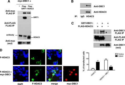
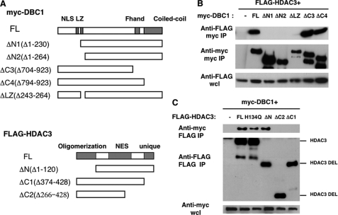
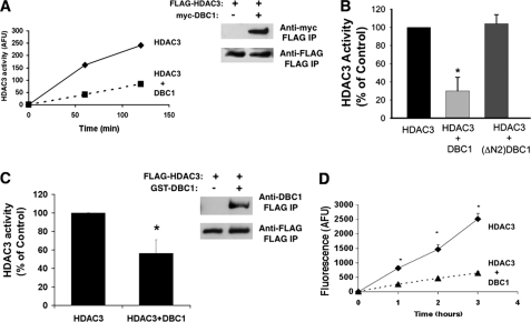
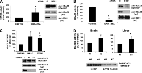
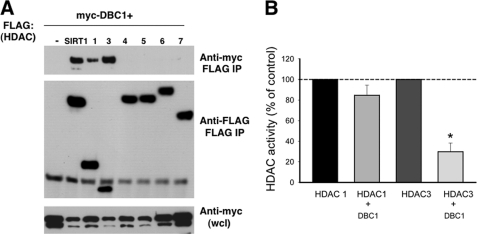
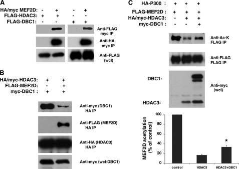
HDAC3 is a member of the class I histone deacetylase family that regulates gene expression by deacetylation of histones and non-histone proteins. HDAC3 activity has been shown to be modulated by interaction with the co-repressors NCoR and SMRT. Here, we present evidence that the nuclear protein DBC1 is an endogenous inhibitor of HDAC3. DBC1 has been previously identified as a regulator of some nuclear receptors, the methyltransferase SUV39H1, and the NAD-dependent deacetylase SIRT1. Furthermore, DBC1 has been shown to influence transcription regulation and apoptosis, and it may also act as a tumor suppressor. We found that DBC1 interacts and specifically inhibits the deacetylase HDAC3. This interaction depends on the N terminus of DBC1 and the C terminus of HDAC3. Expression of DBC1 not only inhibited HDAC3 activity but also altered its subcellular distribution. In addition, knockdown of endogenous DBC1 in cells and knock-out in mouse tissues increased HDAC3 deacetylase activity. Together, these results identify DBC1 as a new regulator of HDAC3 and demonstrate that DBC1 is a negative regulator of two key distinct deacetylases, SIRT1 and HDAC3. These findings may lead to a better understanding of the biological roles of DBC1 and HDAC3 in metabolic diseases and cancer.


Figures

FIGURE 1.
FIGURE 1.
Interaction of DBC1 with HDAC3. A, 293T cells were transfected with expression plasmids for Myc-DBC1 and vector control, FLAG-SIRT1, or FLAG-HDAC3. Proteins were immunoprecipitated (IP) with anti-FLAG antibody and immunoblotted with anti-FLAG and anti-Myc antibodies. wcl, whole cell lysate. B, 293T cell lysates were immunoprecipitated with anti-HDAC3 antibody and immunoblotted with anti-DBC1 and anti-HDAC3 antibodies. C, Sf9 cells were infected with baculovirus expressing GST-DBC1 and FLAG-HDAC3. Cell lysates were immunoprecipitated with anti-FLAG antibody and immunoblotted with anti-FLAG and anti-DBC1 antibodies. D, 293T cells were transfected with FLAG (F)-HDAC3 in the presence and absence of Myc-DBC1. Cells were fixed and stained with anti-FLAG and anti-Myc antibodies and DAPI. The graph shows the percentage of cells that displayed predominantly cytosolic or nuclear HDAC3 staining in the presence and absence of DBC1. About 30 transfected cells were counted for each condition in each experiment. Each experiment was repeated three times. The differences between HDAC3 distribution in the cytosol and nuclei and between HDAC3 alone and in the presence of DBC1 were statistically significant, with p < 0.05 (t test).
FIGURE 2.
FIGURE 2.
The N terminus of DBC1 and the C terminus of HDAC3 are required for the DBC1-HDAC3 interaction. A, shown is a schematic diagram of full-length DBC1 (FL) and deletion mutants of DBC1 and HDAC3. NLS, nuclear localization signal; NES, nuclear export sequence. B, plasmids encoding Myc-tagged full-length DBC1 (FL), deletion mutants, or vector control were transfected in 293T cells along with FLAG-HDAC3. Cell lysates were immunoprecipitated (IP) with anti-Myc antibody and immunoblotted with anti-FLAG and anti-Myc antibodies. wcl, whole cell lysate. C, plasmids encoding FLAG-tagged full-length HDAC3 (FL), mutants, or vector control along with Myc-DBC1 were transfected in 293T cells. Cell lysates were immunoprecipitated with anti-FLAG antibody and immunoblotted with anti-FLAG and anti-Myc antibodies. Representative experiments of at least three independent experiments are shown. DEL, deletion.
FIGURE 3.
FIGURE 3.
DBC1 inhibits the HDAC3 deacetylase activity of transfected cells and recombinant proteins. A, 293T cells were transfected with FLAG-HDAC3 in the presence and absence of Myc-DBC1. HDAC3 activity was measured after immunoprecipitation (IP) of FLAG-HDAC3 with anti-FLAG antibody. One representative experiment of a total of five independent experiments is shown. AFU, arbitrary fluorescence units. B, 293T cells were transfected with FLAG-HDAC3 in the presence and absence of Myc-DBC1 or Myc-DBC1-ΔN2. HDAC3 activity was measured after immunoprecipitation with anti-FLAG antibody. C, Sf9 cells were infected with baculovirus expressing FLAG-HDAC3 in the presence and absence of GST-DBC1. HDAC3 activity was measured after immunoprecipitation of FLAG-HDAC3 with anti-FLAG antibody. The immunoblot shows the levels of HDAC3 and DBC1 in immunoprecipitates in a representative experiment. D, FLAG-HDAC3 and GST-DBC1 were purified from baculovirus. FLAG-HDAC3 activity was measured in the presence and absence of purified DBC1 at three time points. Data in B--D are means ± S.D. (n = 3). *, p < 0.05 (t test).
FIGURE 4.
FIGURE 4.
DBC1 inhibits endogenous HDAC3 deacetylase activity. A and B, HDAC3 activity was measured in HDAC3 immunoprecipitates of A549 cells transfected with control (C), DBC1, and HDAC3 siRNAs. One representative Western blot of A549 cells shows the levels of DBC1 and HDAC3 in cell lysates and immunoprecipitates (IP) used for activity. wcl, whole cell lysate. C, HDAC3 activity was measured in HDAC3 immunoprecipitates of NIH3T3 cells stably expressing a control and two DBC1 shRNA clones. The immunoblot shows one representative experiment blotted with anti-DBC1 and anti-HDAC3 antibodies. Results are means ± S.D. of three independent experiments. *, p < 0.05 (t test). D, HDAC3 activity was measured after immunoprecipitation of HDAC3 from brain homogenates and extracts of liver nuclei from wild-type and DBC1−/− mice. Data show means ± S.D. of three controls and three DBC1−/− mice for liver and six controls and six DBC1−/− mice for brain homogenates. *, p < 0.05 (t test). The immunoblot shows the levels of HDAC3 in three controls and three DBC1−/− mice. KO, knock-out.
FIGURE 5.
FIGURE 5.
DBC1 specifically regulates HDAC3. A, 293T cells were transfected with Myc-DBC1 and several types of FLAG-tagged HDACs. HDACs were immunoprecipitated (IP) with anti-FLAG antibody, and proteins were immunoblotted with anti-Myc and anti-FLAG antibodies. wcl, whole cell lysate. B, HDAC1 and HDAC3 activities were measured after transfection of 293T cells with FLAG-HDAC1 and FLAG-HDAC3 in the presence and absence of Myc-DBC1. A comparison between the percent inhibition of HDAC1 and HDAC3 activities by DBC1 is shown. Values are means ± S.D. (n = 4). *, p < 0.05 (t test). Inhibition of HDAC1 by DBC1 was not statistically significant.
FIGURE 6.
FIGURE 6.
DBC1 inhibits the effect of HDAC3 in MEF2D acetylation. A, HA/Myc-MEF2D was transfected in 293T cells in the presence of FLAG-HDAC3 or FLAG-DBC1. MEF2D was immunoprecipitated (IP) with anti-Myc antibody, and proteins were immunoblotted with anti-FLAG and anti-HA antibodies. B, HA/Myc-HDAC3 and Myc-DBC1 were transfected in 293T cells in the presence and absence of FLAG-MEF2D. Protein lysates were immunoprecipitated with anti-HA antibody and immunoblotted with anti-Myc, anti-HA, and anti-FLAG antibodies. C, FLAG-MEF2D and HA-p300 were transfected in 293T cells in the presence of HA/Myc-HDAC3 and/or Myc-DBC1. Cell lysates were immunoprecipitated with anti-FLAG antibody and immunoblotted with anti-acetyllysine (Anti-Ac-K) and anti-FLAG antibody. Whole cell lysates (wcl) were immunoblotted for HDAC3 and DBC1 with anti-Myc antibody. The graph shows the percent change in the levels of MEF2D acetylation from three independent experiments as determined by densitometry. Values are the mean ± S.D. *, p < 0.05 (t test).

Similar articles

Cited by

References

    1. Haberland M., Montgomery R. L., Olson E. N. (2009) Nat. Rev. Genet. 10, 32–42 - PMC - PubMed
    1. Karagianni P., Wong J. (2007) Oncogene 26, 5439–5449 - PubMed
    1. Villagra A., Sotomayor E. M., Seto E. (2010) Oncogene 29, 157–173 - PubMed
    1. Donmez G., Guarente L. (2010) Aging Cell 9, 285–290 - PubMed
    1. Kim E. J., Um S. J. (2008) BMB Rep. 41, 751–756 - PubMed

Publication types