. 2010 Nov 26;330(6008):1247-51.
doi: 10.1126/science.1189157. Epub 2010 Oct 28.

PML regulates apoptosis at endoplasmic reticulum by modulating calcium release

Affiliations

PML regulates apoptosis at endoplasmic reticulum by modulating calcium release

Carlotta Giorgi et al. Science. .

Abstract

The promyelocytic leukemia (PML) tumor suppressor is a pleiotropic modulator of apoptosis. However, the molecular basis for such a diverse proapoptotic role is currently unknown. We show that extranuclear Pml was specifically enriched at the endoplasmic reticulum (ER) and at the mitochondria-associated membranes, signaling domains involved in ER-to-mitochondria calcium ion (Ca(2+)) transport and in induction of apoptosis. We found Pml in complexes of large molecular size with the inositol 1,4,5-trisphosphate receptor (IP(3)R), protein kinase Akt, and protein phosphatase 2a (PP2a). Pml was essential for Akt- and PP2a-dependent modulation of IP(3)R phosphorylation and in turn for IP(3)R-mediated Ca(2+) release from ER. Our findings provide a mechanistic explanation for the pleiotropic role of Pml in apoptosis and identify a pharmacological target for the modulation of Ca(2+) signals.


Figures

Fig. 1
Fig. 1
Identification of Pml at ER and MAM regions and Ca2+-mediated Pml-dependent cell death. (A) Detection of Pml by immunoblotting in Pml+/+ MEFs fractionation. IP3R, tubulin, proliferating cell nuclear antigen (PCNA), and voltage-dependent anion channel (VDAC) are used as markers. H: homogenate; Mc: crude mitochondria; Mp: pure mitochondria; ER; MAM; C: cytosol; N: nucleus. (B) Immunogold labeling of Pml near the rough ER (r), mitochondria (m), and MAM (arrowheads) in Pml+/+ MEFs. Gold particles (15 nm) are mostly associated with the surface of the ER (7.07 gold particles/µm2) and more occasionally with mitochondrial membranes (3.08 gold particles/µm2) (a and b). Specificity of the antibodies is demonstrated by labeling of nuclear bodies (n) (c). Morphologically identified MAM often demonstrated labeling at contacts between ER and mitochondria [(d) to (g), and arrowheads in insets therein]. Insets correspond to boxed areas. Bar: (a) 360 nm; (b) 340 nm; (c) 370 nm; (d) 188 nm, inset 120 nm; (e) 260 nm, inset 190 nm; (f) 340 nm, inset 180 nm; (g) 280 nm, inset 210 nm. (C) Apoptosis induced by 1 mM H2O2, 15 µM menadione (MEN), 6 µM tunicamycin (TN), 2 µM thapsigargin (TG), or 50 µM etoposide (ETO) in Pml+/+ or Pml−/− MEFs treated for 12 hours. Data represent the mean SD of five independent experiments.
Fig. 2
Fig. 2
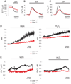
Intracellular Ca2+ homeostasis in Pml+/+ and Pml−/− MEFs. (A to C) ER (A), cytosolic (B), and mitochondrial (C) Ca2+ homeostasis measurements with aequorins. Where indicated, cells were treated with 100 µM ATP. Pml+/+: [Ca2+]ER peak 448 ± 32 µM; [Ca2+]c peak 3.3 ± 0.16 µM; [Ca2+]m peak 138 ± 14 µM. Pml−/−: [Ca2+]ER peak 386 ± 42 µM; [Ca2+]c peak 2.65 ± 0.23 µM; [Ca2+]m peak 78 ± 10 µM. n = 15 samples from five independent experiments, P < 0.01. (D) MEFs loaded with calcium-sensitive fluorescent dye fura-2 were stimulated with menadione (MEN) or H2O2. The kinetic behavior of the [Ca2+]c response is presented as the ratio of fluorescence at 340 nm/380 nm. In these, and other fura-2 experiments, the traces are representative of at least 10 single-cell responses from three independent experiments. (E) Analysis of [Ca2+]m during oxidative stress. Where indicated, cells were stimulated with 30 µM MEN or 2 mM H2O2. n = 10 samples from three independent experiments.
Fig. 3
Fig. 3
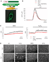
erPML chimera reestablishes the [Ca2+]m and apoptotic responses in Pml−/− MEFs. (A) Schematic map of the erPML chimera and immunofluorescence image, stained with the antibody to PML, of Pml−/− MEFs expressing erPML. (B) erPML reestablishes the agonist-dependent [Ca2+]m response in Pml−/− MEFs ([Ca2+]m peak 135 ± 12 µM) to values comparable to those of Pml+/+ MEFs. (C) Pml−/− and Pml−/− MEFs expressing erPML previously incubated with fura-2 were stimulated with menadione (MEN) or H2O2. (D) Representative microscopic fields of Pml−/− MEFs and Pml−/− expressing erPML before and after treatment with 1 mM H2O2, 15 µM MEN, or 50 µM etoposide (ETO) for 16 hours.
Fig. 4
Fig. 4
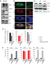
Modulation of [Ca2+]m and apoptotic responses by Pml through Akt- and PP2a-dependent phosphorylation of IP3R3. (A) Coimmunoprecipitation of IP3R3 with Pml, Akt, and PP2a in Pml+/+ MEFs. In the same blot, the levels of p-IP3R3 and pAkt are shown. (B) Localization of Pml (green) and PP2a (red) at ER and MAM sites in Pml+/+ MEFs analyzed by immunofluorescence. FACL [long-chain fatty acid–CoA (coenzyme A) ligase type 4, blue] was used as MAM marker. (C) Pml+/+ MEFs subcellular fractionation and identification of PP2a and Akt at ER and MAM fractions by immunoblot. (D) Effects of okadaic acid (OA, 1 µM for 1 hour) and LY294002 (5 µM for 30 min) on agonist-dependent [Ca2+]m responses in Pml+/+, Pml−/−, and Pml−/− MEFs expressing erPML. [Ca2+]m is represented as a percentage of the peak value of control cells. Representative traces are shown in fig. S15. (E) Quantification of cell survival of Pml+/+, Pml−/−, and Pml−/− MEFs expressing erPML, control (CTR, untreated) and treated first with OA (1 µM for 1 hour) or LY294002 (5 µM for 30 min) and then H2O2 or menadione (MEN) for 16 hours. The data show the percentage of living cells in the whole-cell population negative for annexin-V–fluorescein isothiocyanate and propidium iodide staining, analyzed by flow cytometry. Data show the means SD from three independent experiments.

Comment in

References

    1. Salomoni P, Pandolfi PP. Cell. 2002;108:165. - PubMed
    1. Gurrieri C, et al. J. Natl. Cancer Inst. 2004;96:269. - PubMed
    1. Shen TH, Lin HK, Scaglioni PP, Yung TM, Pandolfi PP. Mol. Cell. 2006;24:331. - PMC - PubMed
    1. Lin HK, Bergmann S, Pandolfi PP. Nature. 2004;431:205. - PubMed
    1. Condemine W, et al. Cancer Res. 2006;66:6192. - PubMed

Publication types

MeSH terms