. 2011 Jan;300(1):G71-81.
doi: 10.1152/ajpgi.00215.2010. Epub 2010 Oct 28.

Role of liver sinusoidal endothelial cells and stabilins in elimination of oxidized low-density lipoproteins

Affiliations

Role of liver sinusoidal endothelial cells and stabilins in elimination of oxidized low-density lipoproteins

Ruomei Li et al. Am J Physiol Gastrointest Liver Physiol. 2011 Jan.

Abstract

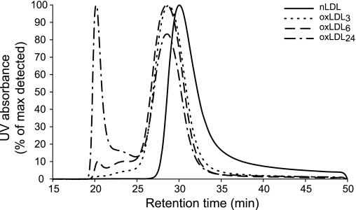
Atherogenesis is associated with elevated levels of low-density lipoprotein (LDL) and its oxidized form (oxLDL) in the blood. The liver is an important scavenger organ for circulating oxLDLs. The present study aimed to examine endocytosis of mildly oxLDL (the major circulating form of oxLDLs) in liver sinusoidal endothelial cells (LSECs) and the involvement of the scavenger receptors stabilin-1 and stabilin-2 in this process. Freshly isolated LSECs, Kupffer cells (KCs), and stabilin-1- and stabilin-2-transfected human embryonic kidney cells were incubated with fluorescently labeled or radiolabeled oxLDLs [oxidized for 3 h (oxLDL(3)), 6 h, or 24 h (oxLDL(24))] to measure endocytosis. The intracellular localization of oxLDLs and stabilins in LSECs was examined by immunofluorescence and immunogold electron microscopy. Whereas oxLDL(24) was endocytosed both by LSECs and KCs, oxLDL(3) (mildly oxLDL) was taken up by LSECs only. The LSEC uptake of oxLDLs was significantly inhibited by the scavenger receptor ligand formaldehyde-treated serum albumin. Uptake of all modified LDLs was high in stabilin-1-transfected cells, whereas stabilin-2-transfected cells preferentially took up oxLDL(24), suggesting that stabilin-1 is a more important receptor for mildly oxLDLs than stabilin-2. Double immunogold labeling experiments in LSECs indicated interactions of stabilin-1 and stabilin-2 with oxLDL(3) on the cell surface, in coated pits, and endocytic vesicles. LSECs but not KCs endocytosed mildly oxLDL. Both stabilin-1 and stabilin-2 were involved in the LSEC endocytosis of oxLDLs, but experiments with stabilin-transfected cells pointed to stabilin-1 as the most important receptor for mildly oxLDL.


Figures

Fig. 1.
Fig. 1.
Size-exclusion chromatography of oxidized low-density lipoprotein (oxLDL). Separation of native LDL (nLDL) and oxLDL with different degrees of oxidation was performed by size-exclusion chromatography using the DIONEX HPLC system and Superose-6 10/300 column (Amersham Pharmacia Biotech). Low-density lipoprotein (LDL) retention time was detected by continuous ultraviolet (UV) absorbance (280 nm) measurement of the eluent. LDL relative molecular mass (Mr) values were calculated from the standard curve generated by plotting the retention time of protein standards against their corresponding Mr. Native LDL eluted as a single peak corresponding to 2,100 kDa. Oxidation induced a shift of the main peak toward a higher-molecular-size region [3,000 kDa for LDL oxidized by copper sulfate (CuSO4) for 3 h (oxLDL3), 3,100 kDa for LDL oxidized by CuSO4 for 6 (oxLDL6) and 24 (oxLDL24) h]. Moreover, with increased oxidation time, the additional peaks of intermediately (13,000 kDa) and heavily (32,000 kDa) aggregated material began to appear and became very pronounced in the sample oxidized for 24 h.
Fig. 2.
Fig. 2.
Confocal microscopy analysis of oxLDL uptake in nonparenchymal liver cells (NPCs). Rat NPCs were incubated with 10 μg/ml of 3,3-dioctadecylindocarbocyanine (DiI)-oxLDL3 (A–C, red fluorescence) or DiI-oxLDL24 (DF, red fluorescence) at 37°C for 30 min; washed and incubated with 0.5 × 10⁶/ml latex beads (ø = 2 μm) for another 30 min; and fixed and immune labeled with antibodies against stabilin-2 (A and D), CD36 (B and E), or the macrophage marker CD163 (C and F). Positive immune labeling was visualized with Alexa-488 goat anti-rabbit/anti-mouse antibodies (green fluorescence), respectively. Cell nuclei were visualized with Draq 5 (blue fluorescence). The images presented are overlays of the triple-fluorescence channels and bright field images. Arrows point to latex beads in Kupffer cells (KCs).
Fig. 3.
Fig. 3.
Endocytosis of oxLDLs in liver sinusoidal endothelial cells (LSECs). A: time course endocytosis in rat LSEC cultures incubated with 0.1 μg/ml of radiolabeled ligands for various time periods at 37°C. Results (means ± SD) are averages of triplicate measurements representing the sum of cell-associated and degraded ligand (details in materials and methods). Corresponding results were found in two other experiments. B: LSEC capacity for endocytosis of oxLDLs. Rat LSEC cultures were incubated with radiolabeled ligand (0.1 μg/ml) alone (control) or together with the indicated amounts of homologous nonlabeled molecules for 1 h at 37°C. The results are means of 4 experiments, representing cells from 4 different animals. Error bars represent SE. C: specificity of endocytosis of oxLDLs in rat LSEC cultures incubated with 0.1 μg/ml of indicated radiolabeled ligands alone (control) or together with excess amounts of nonlabeled formaldehyde-treated serum albumin (FSA) or LDL (100 μg/ml) for 2 h at 37°C. The results shown represent an average of 4 independent experiments. *Statistically significant (P < 0.01) difference in uptake compared with the other two treatments.
Fig. 4.
Fig. 4.
Time course endocytosis of oxLDLs in stabilin-1- and stabilin-2-transfected cells. Confluent nontransfected human embryonic kidney 293 cell (HEK) cultures (A) and cultures of HEK cells stably transfected with mouse stabilin-1 (mS1-HEK, B) and stabilin-2 (mS2-HEK, C) were incubated with 0.1 μg/ml of radiolabeled ligands as indicated at 37°C for various time periods. Results (means ± SD) are averages of triplicate measurements representing the sum of cell-associated and degraded ligand (details in materials and methods). Corresponding results were found in two other experiments.
Fig. 5.
Fig. 5.
Specificity of endocytosis of oxLDLs in stabilin-1- and stabilin-2-transfected cells. Confluent mS1-HEK cultures (A) and mS2-HEK cultures (B) were incubated with 0.1 μg/ml of the indicated radiolabeled ligands alone (control) or together with excess amounts of nonlabeled FSA or LDL (100 μg/ml) for 2 h at 37°C. The results presented are an average of three independent experiments. *Statistically significant (P < 0.01) difference in uptake compared with the other two treatments. **Statistically significant (P < 0.01) difference in uptake compared with control only.
Fig. 6.
Fig. 6.
Confocal microscopy analysis of stabilin-1 and oxLDL colocalization in LSECs. A–C: pulse-chase experiment. Rat LSEC cultures were incubated with 40 μg/ml of DiI-oxLDL3 at 4°C for 1 h and then washed and incubated in medium only at 37°C for 20 min. D–F: cells were incubated with 10 μg/ml of DiI-oxLDL3 at 37°C for 10 min and then incubated in the presence of 10 μM monensin for another 50 min to inhibit intracellular traffic and recycling of receptor. The cells were immune labeled with anti-human S1 antiserum and visualized with Alexa-488 goat anti-rabbit antibody. Arrows point to colocalized DiI-oxLDL3 (red fluorescence) and stabilin-1 (green fluorescence) labeling in corresponding channels. Cell nuclei are visualized with Draq 5 (blue fluorescence) in the overlays.
Fig. 7.
Fig. 7.
Confocal microscopy analysis of stabilin-2 and oxLDL colocalization in LSECs. A–C: pulse-chase experiment. Rat LSEC cultures were incubated with 40 μg/ml of DiI-oxLDL3 at 4°C for 1 h and then washed and incubated in medium only at 37°C for 20 min. D–F: rat LSEC cultures were incubated with 10 μg/ml of DiI-oxLDL3 at 37°C for 10 min and then incubated in the presence of 10 μM monensin for another 50 min. The cells were fixed and immune-labeled with anti-rat S2 antiserum and visualized with Alexa-488 goat anti-rabbit antibody. Arrows point to colocalized DiI-oxLDL3 (red) and stabilin-2 (green) labeling in corresponding channels, and cell nuclei are visualized with Draq 5 (blue fluorescence).
Fig. 8.
Fig. 8.
Double immunogold labeling for stabilin-1/-2 and oxLDL3 in LSECs. Rat LSECs were incubated with 40 μg/ml oxLDL3 for 15 min at 37°C, fixed, and processed for immunogold labeling as described in materials and methods. Arrows point to close colocalization of small and large gold particles, indicating direct receptor-ligand interaction. Arrowheads point to the plasma membrane. N, cell nucleus; CP, coated pits. Scale bars = 200 nm. A and B: close colocalization of oxLDL3 (5 nm gold) and stabilin-1 (10 nm gold) is seen in an endocytic vesicle and at the plasma membrane surface in A and in a CP in B. CE: close colocalization of stabilin-2 (5 nm gold) and oxLDL3 (10 nm gold) is seen in larger endosomes (C), coated vesicles (D), and in CP (E).

References

    1. Adachi H, Tsujimoto M. FEEL-1, a novel scavenger receptor with in vitro bacteria-binding and angiogenesis-modulating activities. J Biol Chem 277: 34264–34270, 2002 - PubMed
    1. Avogaro P, Bon GB, Cazzolato G. Presence of a modified low density lipoprotein in humans. Arteriosclerosis 8: 79–87, 1988 - PubMed
    1. Berliner JA, Territo MC, Sevanian A, Ramin S, Kim JA, Bamshad B, Esterson M, Fogelman AM. Minimally modified low density lipoprotein stimulates monocyte endothelial interactions. J Clin Invest 85: 1260–1266, 1990 - PMC - PubMed
    1. Blomhoff R, Eskild W, Berg T. Endocytosis of formaldehyde-treated serum albumin via scavenger pathway in liver endothelial cells. Biochem J 218: 81–86, 1984 - PMC - PubMed
    1. Bourret G, Brodeur MR, Luangrath V, Lapointe J, Falstrault L, Brissette L. In vivo cholesteryl ester selective uptake of mildly and standardly oxidized LDL occurs by both parenchymal and nonparenchymal mouse hepatic cells but SR-BI is only responsible for standardly oxidized LDL selective uptake by nonparenchymal cells. Int J Biochem Cell Biol 38: 1160–1170, 2006 - PubMed

Publication types

LinkOut - more resources